Biomedical Engineering Reference
In-Depth Information
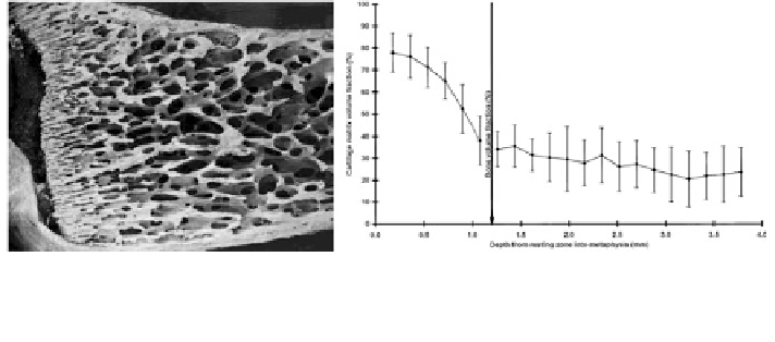
Fig. 7 Scanning electron microscope image of endochondral bone in human neonate rib (left),
and graph showing change in cartilage volume fraction and bone volume fraction (y-axis) versus
distance from the resting zone to the mid-metaphysis (x-axis) (right)[
33
] with permission
5 Trabecular Bone Structure in the Young
Although there have been relatively few studies describing trabecular bone
structure in development and adolescence, the morphological events in bone
growth are well described [
14
,
33
,
42
] (Fig.
7
) and the rate of acquisition of
bone mass has been shown to take place relatively slowly until puberty when the
rate significantly increases [
45
]. The velocity of bone growth is different between
boys and girls, reflecting differences in onset of puberty and in response to dif-
ferences in musculature and while there is convergence in growth rates towards
adulthood,
clear
sex-related
size
differences
in
the
skeleton
are
maintained
throughout life [
45
,
109
].
There is less information as to the relative importance of the genetic template
for bone structure versus the effects of environmental factors, i.e., nature versus
nurture. In the neonate growth plate histological studies clearly show that the
spatial arrangement of the columnar hypertrophic chondrocytes give rise to pri-
mary spongiosa and hence the secondary trabeculae [
14
,
42
]. Whether the quality
of adult trabecular bone structure is determined or influenced at this early stage has
been hypothesized [
33
], although the ability of bone to adapt to the prevailing
mechanical environment shows that genetic influence cannot fully determine an
adult's bone microarchitecture.
There is a peak in fracture incidence in the young around the time of puberty,
which in girls is approximately 11.5-12.5 years, and in boys is approximately
13.5-14.5 years [
45
]. These fractures are commonly associated with moderate
trauma, with the majority occurring in the distal radius. While trabecular bone
structure has not been directly implicated as contributing to susceptibility to
fracture it has been shown that during adolescence there is a transient increase in
longitudinal and circumferential growth of the cortex before there is a corre-
sponding increase in bone mass through thickening of the cortex and consolidation
of the trabecular bone structure within the medullary space [
45
,
60
].
Search WWH ::

Custom Search