Biomedical Engineering Reference
In-Depth Information
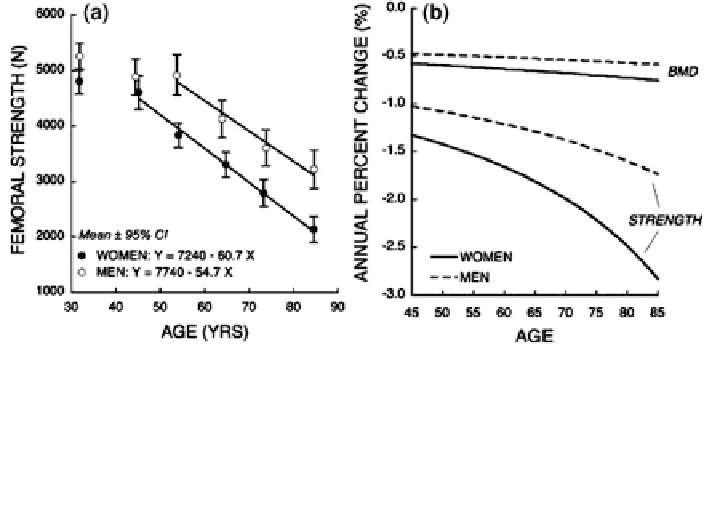
Fig. 3 Change in estimated femoral strength with age (from Keaveny et al. [
60
], with
permission). Strength was predicted from finite element models simulating impact from a
sideways fall on the proximal femur. a Predicted strength declines after age 50 in women and
after age 60 in men. When presented as mean values for each decade, the rates of decline are
linear and appear similar in women and men. b The annualized change in predicted strength and
areal BMD reveal much greater rates of decline in strength than BMD, and also reveal
accelerating percentage declines in strength with increasing age
estimated strength of the distal tibia using HR-pQCT-based FEA in 442 women
and 202 men (20-99 years). In women, strength declined approx. -6 %/decade
versus -3 %/decade in men. Thus, although accessibility to pQCT imaging makes
it an attractive site for study, the distal tibia does not appear to have the dramatic
age-related declines in strength that occur at the proximal femur.
3.2.2 Upper Extremity: Distal Radius
Several studies have reported age-related decreases in bone strength of the distal
radius based either on mechanical testing or FEA. Bonel et al. [
64
] performed
compression tests on forelimbs from 25 female and 24 male cadavers (57-100 years).
The majority of specimens (73 %) failed by fracture of the distal radius. In this group,
bones from females were 30 % weaker than from males, which corresponded to a
25 % smaller bone size (TA). Both sexes had evidence of age-related decline in bone
strength, although the change in females was significant (-14.4 %/decade;
p \ 0.05) whereas the change in males was not (-6.6 %/decade; p [ 0.05).
Mueller et al. [
52
] examined the distal radius from 50 female (mean age 82 yrs)
and 50 male (mean age 80 yrs) cadavers using HR-pQCT and mechanical testing that
produced Colles-type fractures. They scanned the distal 20 % of the bone, but noted
that failure force correlated most strongly with trabecular morphology in the most
distal 4 % (''region 1'' in their paper); results reviewed here are from that region.
Bones from women were smaller, less dense and less strong than men. Total tissue
Search WWH ::

Custom Search