Biomedical Engineering Reference
In-Depth Information
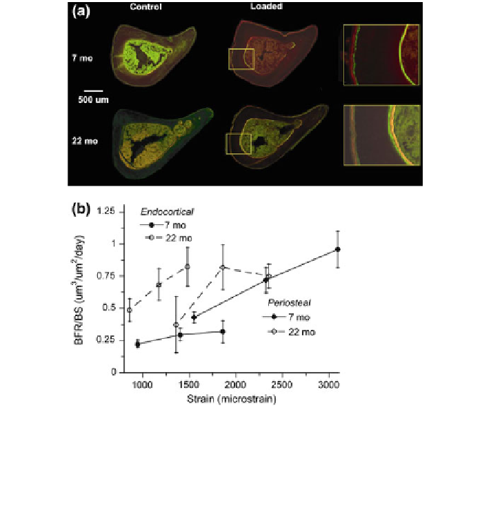
Fig. 2 a Fluorescent photomicrographs of mid-diaphyseal tibial sections from loaded and
control tibias of 22- and 7-months mice. Samples were collected on day 11 following tibial
compression on days 1-5, and fluorochrome labeling on days 5 (green) and 10 (red). An increase
in endocortical and periosteal labeled surface is evident in loaded tibias of both ages compared to
controls. b Bone formation rate measured from tibial sections (n = 8-13 mice/group) from
the same study. There is no loss of mechanoresponsiveness in the old mice. (From Brodt and
Silva [
57
])
that this age was most responsive to loading. Unexpectedly, trabecular bone
volume fraction (BV/TV) was reduced in loaded limbs compared to controls in
4, 7 and 12 month groups, and was unchanged with loading in the 2 month group.
We concluded that, at cortical sites mechanical loading can overcome the normal,
age-related decline in bone formation in mice, with some evidence that the
young-adult skeleton (4 months) is more responsive than the mature to middle-
aged skeleton (7-12 months).
Lynch et al. recently used the axial tibial loading model in two separate studies
to compare bone adaptive responses in young, growing (2 months) versus mature,
adult (6 months) mice [
59
,
60
]. Female C57Bl/6 mice were subjected to 2 weeks
of daily loading (1200 or 2200 le periosteal; 1200 cycles/day, 5 days/week)
and morphology of cortical and trabecular bone was assessed by post hoc
microCT. Comparisons between ages were made challenging because of different
Search WWH ::

Custom Search