Biology Reference
In-Depth Information
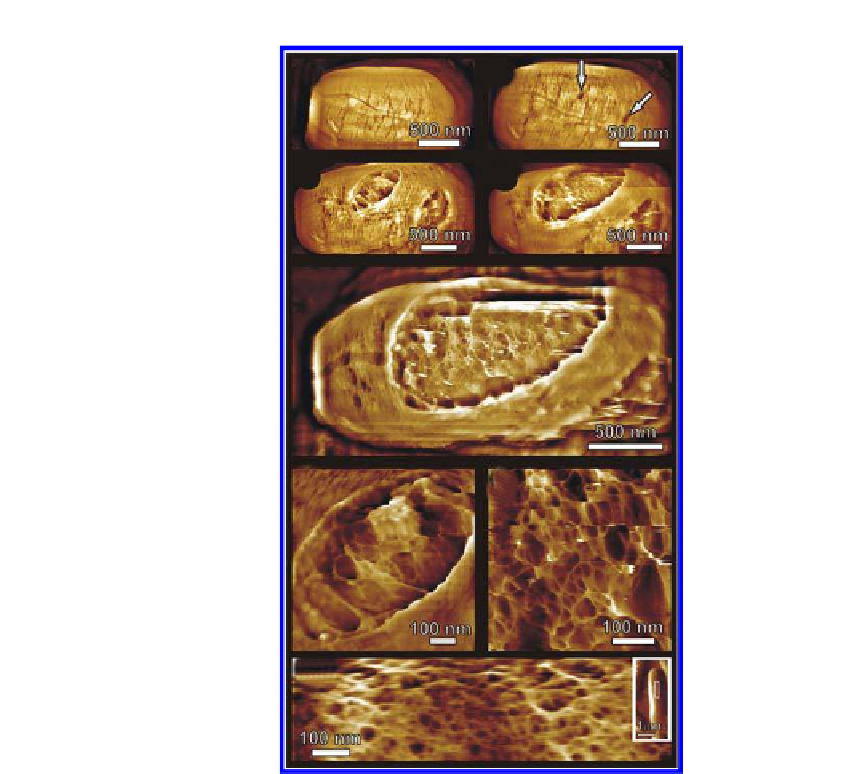
(a)
(b)
(c)
(d)
(e)
(f)
(g)
(h)
Figure 4.10.
Emergence of vegetative cells. (a-g) Series of AFM height images
showing 60-70 nm deep apertures in the rodlet layer (indicated with arrows in (b))
that gradually enlarged (c-d) and subsequently eroded the entire spore coat (e). Germ
cells emerged from these apertures. (e) Prior to germ emergence from the spore coat,
the peptidoglycan cell wall structure was evident. (f ) At an early stage of emergence,
the cell wall was still partly covered by spore remnants, while (g) immediately prior
to cell emergence, the cell wall was free of spore integument debris. The germ cell
surface contained 1-6 nm ibres forming a ibrous network enclosing pores of 5-100
nm. Images in (a-g) were collected on the same spore as those shown in
Figure 4.8e,f.
Elapsed germination time (in hr:min) was (a) 3:40, (b) 5:45, (c) 7:05, (d) 7:30, (e)
7:45, (f ) 7:15, (g) 7:50. (h) In separate experiments, cultured vegetative
B. atrophaeus
cells were adhered to gelatin surfaces and imaged in water. AFM height images show
a slightly denser, similar ibrous network compared with the germ cell network
structure (g), with 5-50 nm pores. In the inset, the imaged part (h) of the entire cell is
indicated with a white rectangle. Images reproduced, with permission from Ref. 7. ©
(2007) National Academy of Sciences, USA.


Search WWH ::

Custom Search