Biology Reference
In-Depth Information
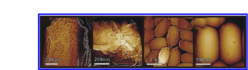
(a)
(b)
(c)
(d)
Figure 4.4.
AFM images of
B. subtilis
spores of different strains. The spores analyzed
were wild type (a),
cotE
(b),
gerE
(c) and
cotEgerE
(d). Images reproduced, with
permission from Refs. 11 and 12. © (2008) American Society for Microbiology.
Proper assembly of a multilayer spore coat of
Bacillus
spores is
dependent on a number of coat proteins.
Loss of any of those proteins could
alter signiicantly the mechanisms of the spore coat assembly and the inal
spore coat structure. Indeed, as demonstrated in
Fig. 4.4
, deletion of a single
spore coat protein could result in pronounced changes in the spore coat
architecture.
1
11,12
Thus, the AFM analysis demonstrated that intact wild-type
B.
subtilis
spores are completely or partially covered by a thin amorphous layer
B.
atrophaeus
is a major spore coat morphogenetic
protein, and in its absence, the outer coat fails to assemble properly.
rodlet spore coat layer.
6
CotE
18
Indeed,
we have demonstrated that for most
spores the outermost structure is
which likely correspond to the inner coat layers.
Furthermore, surfaces of some
cotE
mutant spores exhibit patches or large
regions covering the spore of a hexagonal crystalline layer (located between
of
cotE
spores were found to lack completely both amorphous and rodlet
structures, being encased in several inner spore coat layers.
gerE
7
Finally, spores
lacking both
spores) were found to lack all
outer and inner coat structures, with the spore cortex being the outermost
structure.
CotE
and
gerE
proteins (
cotEgerE
12
Our recent comprehensive analysis of a wide range of
B. subtilis
mutants, which lack various spore coat proteins,
has provided improved
understanding of the spore coat architecture, assembly and function of coat
proteins.
To observe the structure of the
19
spore coat beneath the
amorphous shell, we developed procedures to remove the shells by chemical
treatment with various reducing agents and detergents or by physical
treatment using a French Press.
C. novyi-NT
When either French Press or chemical
treatments were used, the majority of the exposed spore coat surface is
8


Search WWH ::

Custom Search