Biology Reference
In-Depth Information
“material” with repeated scanning. This observation has been described for
both bacteria and for bacterial spores when sample height exceeded 1 μm.
47
Velegol
observed that the orientation of the “material” was the same
despite changes in the scan direction, and they noticed a consistent angle of
±27° to the scan direction, thereby showing that the “material” really was an
imaging artifact.
47
et al.
(a)
(b)
(c)
(d)
(e)
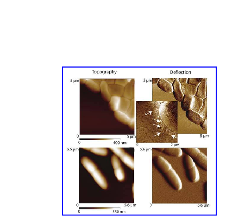
Figure 3.4.
Contact mode AFM images in air (a-c) and in water (d-e) show differences
in resolution of ine structure. Panels (a-b), respectively, are topographic and
delection images while (c) is a friction image of enteroaggregative
E. coli
042. The
white arrows in the friction image point to imbriae, which are clearly visible in image
(c). In (d) and (e), images of
P. aeruginosa
PA14 taken in water are shown. The reduced
level of details showing imbriae and cellular surface structure is especially obvious
when comparing the delection mode images taken in air and water.
The shape of the artifact is explained as being a combination of the
object being scanned, the geometry of the tip, the angle of tip tilt and the
scan direction.
47
The choice of cantilever is an important one because of the
available variations in material, size, shape and tip geometry. Collectively,
these features determine the mechanical forces imposed on the sample
during imaging. Commercially available silicon cantilevers are stiffer than
their silicon nitride counterparts. High aspect ratio tips which have a smaller
radius of curvature are better for imaging rigid samples with tall features,
but they are costlier and more fragile than low aspect ratio tips. On the other


Search WWH ::

Custom Search