Biology Reference
In-Depth Information
exosomes considered as potential non-invasive biomarker resource for
oral cancer
19
have been studied recently using AFM.
25
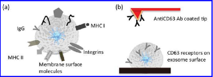
Single exosomes
vesicle ultrastructure, quantitative surface molecular constitution
and nanomechanical characteristics of exosomes may be helpful for
understanding the role of exosomes in intercellular communication
and delivery of genetic components through the extracellular domain
(
Fig. 20.4a
)
.
AFM has developed as a useful single-molecule tool for sensing and
mapping molecular recognition interactions on biological cell interfaces.
26,27
Cell type-speciic markers such as CD63 receptors on individual exosomes can
be analysed using force spectroscopy. Force spectroscopy relies on measuring
the interaction force with piconewton sensitivity as the tip is pushed towards
the sample and retracts from it in the
direction. The force is monitored
by measuring the delection (vertical bending) of the cantilever. Measuring
molecular receptors on the exosome surface requires recording force curves
between the modiied tips (antiCD63 antibody) and the exosomes surface.
At large tip-sample separation distances, the force experienced by the tip
is zero. As the tip approaches the surface, the cantilever may bend upwards
owing to repulsive forces until the tip jumps into contact with the exosome
surface (
Fig. 20.4b
)
. Upon retracting the tip from the surface, in the event of
successful binding of the antiCD63 antibody to the complementary receptors
on the vesicle surface, the curve shows an unbinding event calculated as the
adhesion “pull-off ” force. The rupture force represents the unbinding force
between complementary antiCD63 IgG receptors and ligand molecules borne
on the vesicle outer membrane. The recognition of single receptor molecules
on biological luid-derived exosomes, such as saliva, can potentially detect
surface tumour-antigen-enriched cancer exosomes, and thereby enable early
cancer diagnosis where conventional methods may prove ineffective because
of sensitivity limitations.
z
Figure 20.4.
(a) Schematic showing exosome vesicle and surface receptors. (b)
Schematic of receptor recognition spectroscopy via adhesion force measurements
between AntiCD63 IgG-functionalized AFM tips and exosome surface.


Search WWH ::

Custom Search