Biology Reference
In-Depth Information
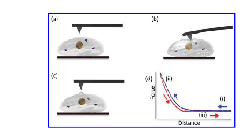
Figure 20.3.
Schematic of an AFM probing a cell surface (a) AFM tip approaching
cell surface, (b) indented into cell surface and (c) retracted from cell surface.
(d) Typical force-displacement curve ((i-iii) correspond to the positions described
earlier), recorded as the “approach” and “retract” curves of the cantilever as it moves
towards and away from the surface. The force acting on the cantilever is recorded as
a function of the piezoelectric crystal displacement. Mechanical properties, such as
the Young's modulus (E) or cell stiffness, can be calculated from force curves using a
Hertz model.
by recording the delection (vertical bending) of the cantilever. As described
earlier, the cantilever delection is usually detected by a laser beam focused
on the free end of the cantilever and relected into a photodiode; this
delection is directly proportional to the force. Force-displacement curves
are obtained by monitoring the delection of the cantilever (
Fig. 20.3d
)
. The
microcantilever-based system allows us to probe the local Young's modulus
(E) or “stiffness” of living cells, performs force spectroscopy measurements
with piconewton resolution and provides a sensor to record
measurements of the cell wall at sub-nanometre resolution. In particular,
AFM is a key tool in acquiring kinetic information, and real-time signals of
living cells, and is capable of offering
in vivo
single-cell diagnostics. AFM
measurements provide a greater understanding of structure, function and
relationships of biological macromolecules, thus generating characteristics
inherent to speciic biological cells.
15
These emerging concepts aid in the
development of new types of nanomechanical sensors, which may contribute
signiicantly to the understanding of changes in cytoarchitecture, which
are characteristic of cellular de-differentiation, malignant transformation,
growth activation, cell motility and disease states.
in vivo


Search WWH ::

Custom Search