Biology Reference
In-Depth Information
of the cell can be determined by recording the cantilever delection as it
decreases during cell relaxation and internal remodelling of the cytoskeleton
(
Fig. 18.8
). To qualitatively visualize the deformation and relaxation processes
in the actin, MT and IF cytoarchitecture we employed cells (human foreskin
ibroblasts cultured as described in section 18.3.1) transiently expressing
GFP-actin, GFP-tubulin and GFP-vimentin, respectively (
Fig. 18.9
).
(a)
(b)
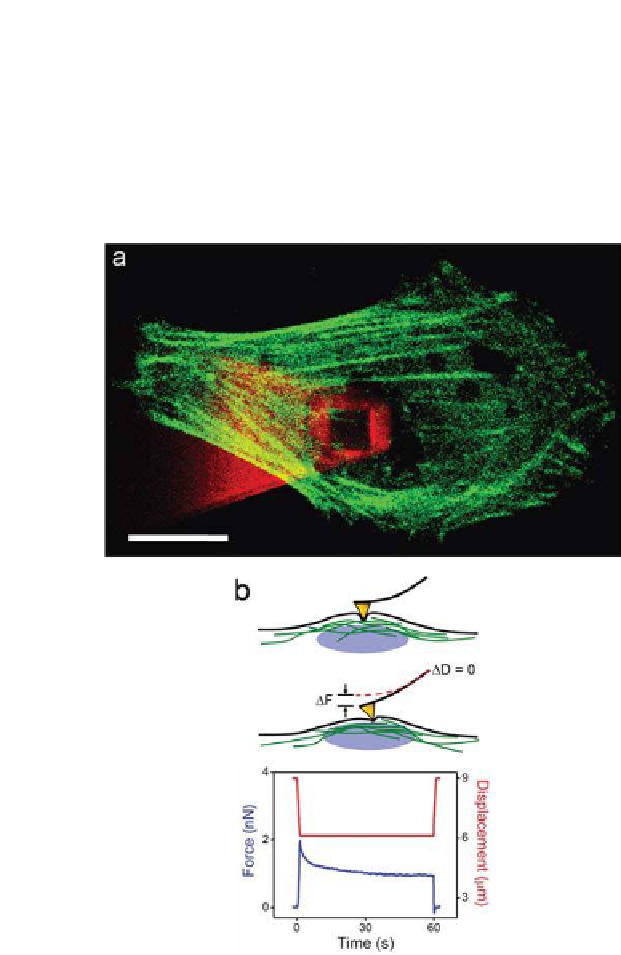
Figure 18.8.
(a) LSCM image of a cell transiently expressing GFP-actin (green).
3
The
AFM tip can be visualized by capturing the autoluorescence resulting from excitation
with a 405 nm diode laser (scale bar = 10
μ
m). b) Stress-relaxation experiments can
be performed in which the AFM tip is brought into contact with the cell at a speciic
setpoint force. The cells are then allowed to relax while the cantilever delection
is monitored as a function of time. This type of measurement yields the cellular
viscosity. Confocal stacks acquired immediately before and after the experiment
allow one to directly visualize cytoskeletal deformation in response to local forces.


Search WWH ::

Custom Search