Biology Reference
In-Depth Information
Previously we have shown that nuclei and cytoskeleton deformations
were observed following local AFM indentation.
72
Here, we review our work
that demonstrates the effect of instantaneous displacement of luorescently
labelled mitochondria upon the static application of force with the AFM.
18,73
Mitochondria form dense three-dimensional (3D) networks around the
nucleus and become lattened and more sparsely distributed at the edges
of the cell. We examined how locally applied forces above the nucleus are
physically transmitted over long distances to the cell edge. It was impossible
to distinguish and separate two-dimensional (2D) versus 3D movement of
mitochondria around the nucleus in response to applied force from the AFM
tip because of the thickness of the cell. Therefore, we limited our analysis to
the cell edge. In these regions, the cell is very lat, as little as 200 nm thick,
and mitochondria are assumed to move perpendicular to the normal force
delivered by the AFM tip over the nucleus, enabling accurate measurement
of physical force transduction from the AFM tip. Furthermore, individual
mitochondria can be resolved much more clearly in these regions, allowing
for accurate image registration and tracking analysis.
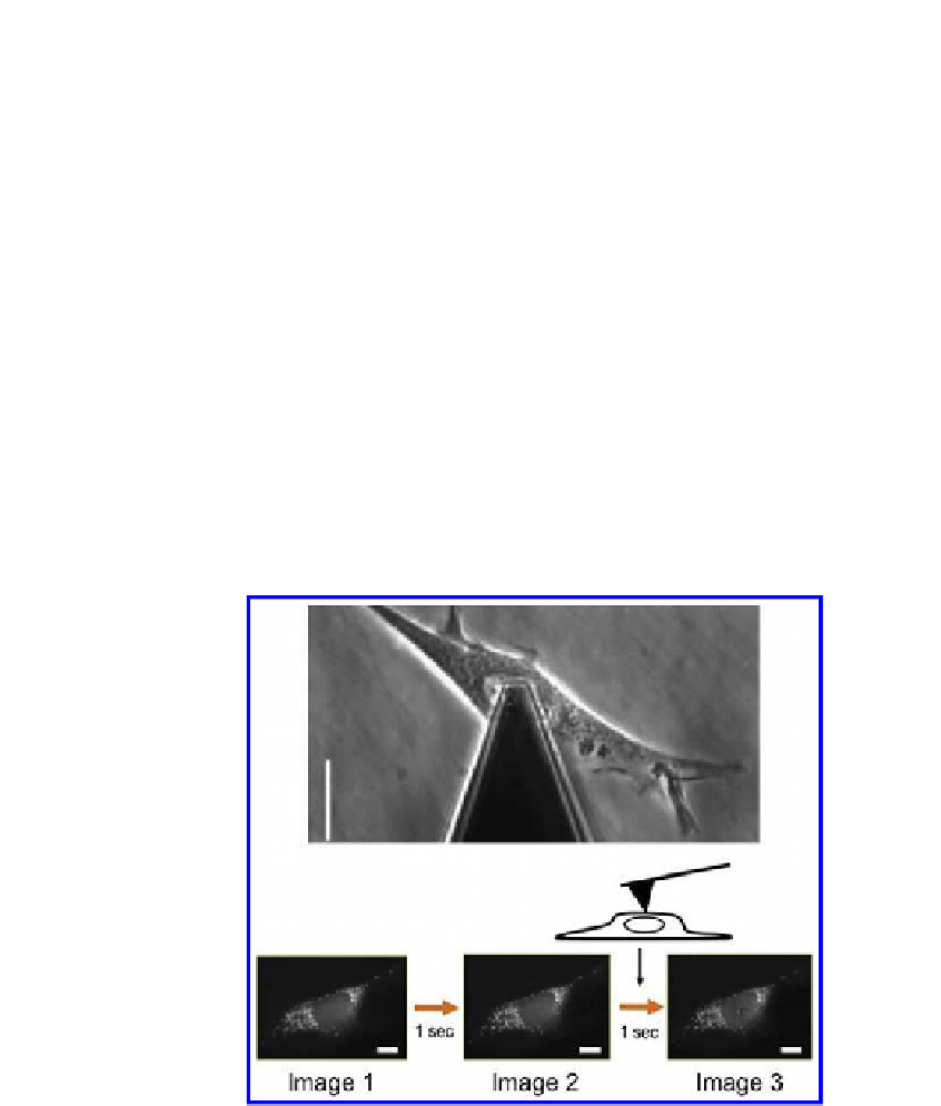
Figure 18.2.
A typical phase-contrast image of the AFM tip and a living cell (scale
bar = 10 μm).
18
A sequence of images is then acquired at 1 second intervals. Three
images were picked for analysis: 2 images taken prior to AFM indentation (images 1
and 2) and the one image that followed the indentation (image 3). Changes between
image 1 and 2 relect basal mitochondrial movement, while changes between image
2 and image 3 relect the force-induced movement resulting from AFM indentation.


Search WWH ::

Custom Search