Biology Reference
In-Depth Information
of lipid rafts differed from that of the surrounding membrane, Roduit
et al.
39
recorded force-volume images of living neurones using an aerolysin-coated
AFM tip. Aerolysin is known to interact with molecular domains that are
speciically expressed within the lipid rafts. Consequently, during scanning
with an aerolysin -coated AFM tip, it should be possible to detect speciic
interactions with the lipid rafts. By comparing the stiffness of the regions in
which a speciic interaction occurred with the stiffness of the surrounding
domains, the authors were able to demonstrate that the lipid rafts were about
Finally, it has recently become possible to glean information concerning
the mechanical properties of subcellular components within living cells from
AFM imaging.
For this purpose, a more sophisticated analysis of the FD
curves is called for. Instead of itting the entire FD curve to the Hertz model,
small segments are individually analyzed. The stiffness of each segment is
then displayed as a colour-coded volume within a three-dimensional matrix.
Using this technique, it is possible to distinguish cytoskeletal components
40
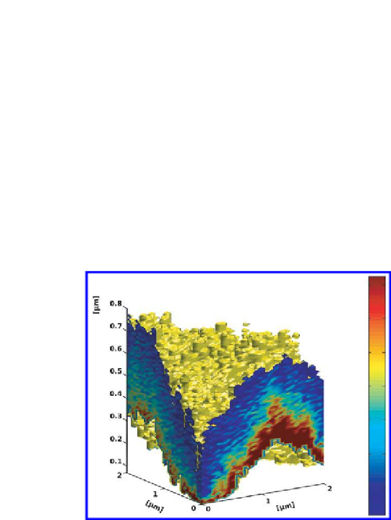
Figure 16.8.
Stiffness tomography of an axon. This imaging mode reveals structures
hidden in the bulk of the sample. The stiffness of the intracellular structures is colour-
coded (blue: soft; red: hard). Scanning distance: 2
M
m (kindly provided by Dr. C.
Roduit).


Search WWH ::

Custom Search