Biology Reference
In-Depth Information
10.3.3.1 Detachment force
F
D
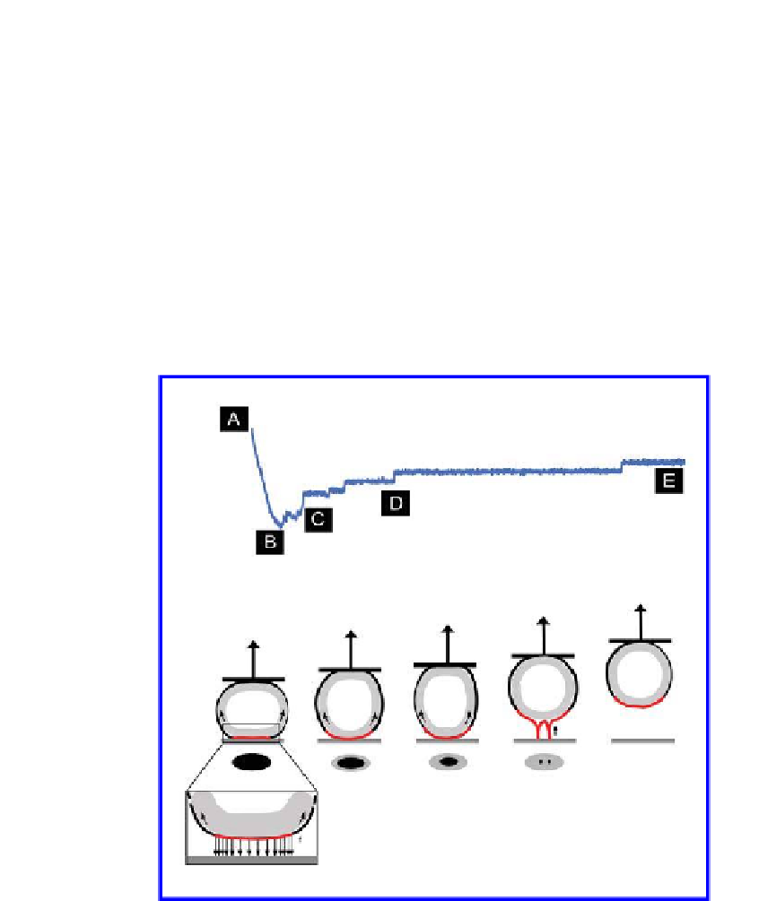
it is usually observed that a circular area approximates the contact zone
between cell and substrate. During the initial detachment phase, bonds in
the outer contact zone are predominantly stressed. The cell is stretched until
a maximal force is reached. Upon bond failure, the contact zone shrinks.
Assuming a homogenous distribution of receptors over the contact zone, more
bonds per radial section will have formed at the periphery of the contact zone
than in the inner region. Consequently, a maximal force is detected before the
bonds at the periphery begin to rupture. Subsequently, the force decreases
quickly since the applied force load is shared by fewer receptors in the inner
(a)
(b)
(c)
(d)
(e)
Figure 10.5.
Schematic representation of the cell detachment process. The
detachment process of a cell can be separated into different phases. (a) The cell is
in contact with the substrate. In the contact zone (red) adhesive interactions are
established. (b,c) During cell detachment, the established interactions (speciic and
non-speciic) bonds rupture and the contact zone shrinks. When the cell body is
separated from the substrate, membrane tethers (nanotubes) link cell and substrate
(d) until the cell is fully detached from the substrate (e).


Search WWH ::

Custom Search