Biology Reference
In-Depth Information
When using a functionalized AFM tip and reduced contact times
and contact forces between tip and cell surface, the binding probability of
the probing tip with the cell surface is rather low. In this case, single binding
events dominate, and the assay may be rather related to single-molecule
force spectroscopy approaches such as discussed in
Chapters 11, 12
and
15
.
In the majority of cell-surface interaction studies, the irst setup (
Fig. 10.1a
)
has been applied and will therefore be detailed.
32
(a)
(b)
(c)
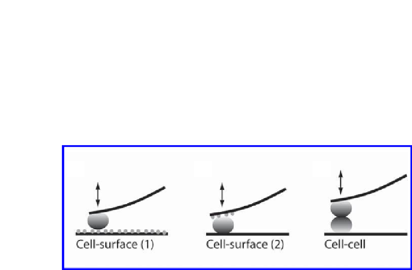
Figure 10.1.
SCFS setups to measure cellular interactions with adhesive substrates.
(a) A single cell is immobilized to a tip-less AFM cantilever, and the adhesion of the
cell to a substrate is probed. (b) A cell, attached to a supporting surface, is probed with
a ligand-coated cantilever. (c) To quantify cell-cell adhesion, a cell immobilized to a
supporting surface is probed with another cell attached to a cantilever.
10.3.1 Converng a Living Cell into a Probe
To attach a living cell to the cantilever, the cantilever surface has to be func-
tionalized with an adhesive substrate. For the immobilization of eukaryotic
cells, concanavalin A, a lectin that binds mannose residues of glycoproteins on
the cell surface,
33
is frequently used.
30,34-39
For certain cell types, e.g., T cells,
the use of concanavalin A may be problematic since it can lead to cell activa-
tion.
40
Alternatively, wheat germ agglutinin,
41
ECM proteins,
42,43
polyphenolic
proteins extracted from marine mussels
44,45
or antibodies
42
can be used to at-
tach different cell types to the AFM cantilever. In other studies, cells were bio-
tinylated and attached to a streptavidin-modiied cantilever,
46
or cells were
directly grown on the cantilever.
31
To attach a cell to a functionalized cantilever, suspended cells are added
into a temperature-controlled luid chamber. Cantilever and cell are visualized
by light microscopy and positioned relative to each other. Then, the cantilever
is lowered onto a single cell, gently pressed on it and withdrawn to capture


Search WWH ::

Custom Search