Biology Reference
In-Depth Information
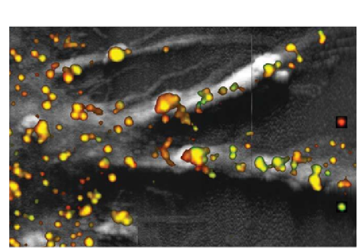
Figure 9.5.
Combined topography (gray) and NSOM image (colour) of the pathogen
recognition receptor DC-SIGN expressed on immature dendritic cells. Spots have
different size and intensity relecting the nanocluster organization of DC-SIGN. Image
adapted with permission from Ref. 60. © (2007) Wiley-VHC.
In the case of members of the epidermal growth factor (EGF) receptor
tyrosine kinase family, clustering is thought to have an adverse effect. Some
EGFs, like the erbB2 receptor, are found to be over-expressed in breast
cancerous cells. It is thought that this over-expression leads to cluster
formation causing the highly oncogenic activation of very potent kinase
activity. Indeed, by applying NSOM in air, the clustering behaviour of EGF
receptors was found to be associated with the activation state of the cell.
62
Additionally, it was found that EGF cluster sizes increased if the quiescent cells
were treated with EGF activators to the same extend as cells over-expressing
these EGFs.
62
Since activation of the EGF signalling pathways requires
extensive interaction between individual members of the EGF family, it is
likely that concentrating one of these EGF receptors in clusters increases the
likelihood of co-clustering of other EGF members. This co-clustering would
then subsequently increase the EGF signalling eficiency. In other words, a
higher local concentration will decrease the lag time for direct inter-receptor
contact.
Cell-signalling events commonly involve a multitude of spatially
segregated proteins and lipids. As such, standard confocal microscopy
studies in biology usually involve multiple colours corresponding to multiple
speciically labelled proteins. However, inherent to all lens-based techniques


Search WWH ::

Custom Search