Biology Reference
In-Depth Information
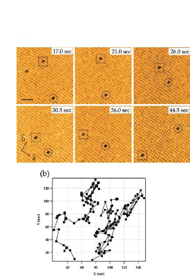
two axes of the crystalline lattice. These defects have larger mobility along
the
b
-axis than along the
a
-axis.
(a)
(b)
Figure 8.2.
Migration of monovacancy defects in streptavidin 2D crystal. (a) High-
speed AFM images of streptavidin 2D crystal and monovacancy defects therein. The
monovacancy defects are enclosed by dashed squares and circles. The directions of
the lattice vectors of the crystal are also indicated. Successive images were obtained
at an imaging rate of 0.5 s/frame with a scan area of 150
s
150 nm
2
. (b) Trajectories
of individual monovacancy defects. Closed squares and circles correspond to defects
indicated by open squares and circles shown in (a), respectively.


Search WWH ::

Custom Search