Biomedical Engineering Reference
In-Depth Information
d
n
4
y
3
n
g
|
8
Figure 7.3
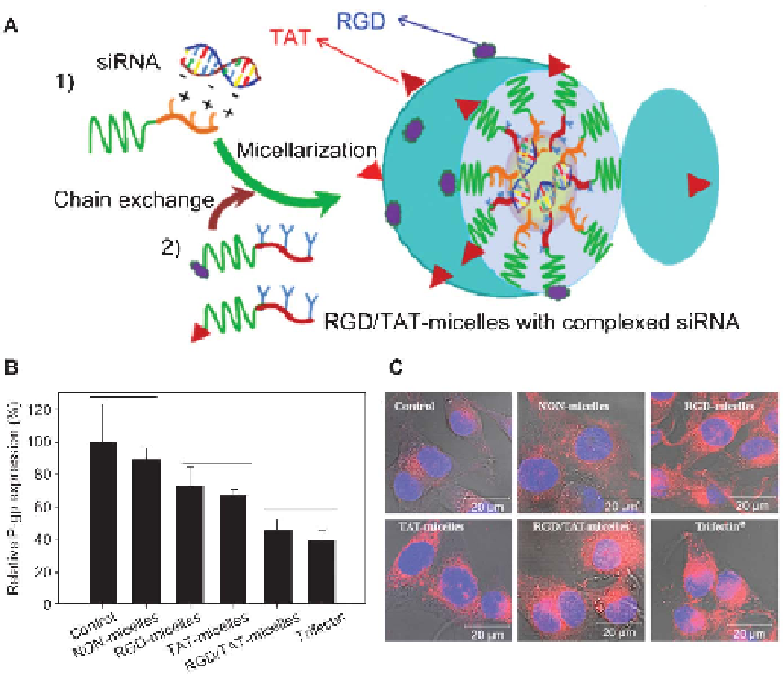
(A) Schematic illustration of RGD4C and/or TAT polymeric micellar
siRNA complex formation. (B) P-gp expression by flow cytometry by
comparing the P-gp related fluorescence intensity to untreated controls
after 48 h of incubation. (C) Reversal of resistance to DOX in resistant
MDA435/LCC6 cells after transfection with mdr1 siRNA formulations.
After transfection, the cells were exposed to free DOX (5 mg mL
21
) and
assessed for DOX cellular accumulation and distribution by fluorescence
microscopy. (Adapted from Xiong and Uludag
69
with permission from
Elsevier.)
and protein levels, leading to increased cellular accumulation of doxorubicin
(DOX) in the cytoplasm and nucleus. RGD/TAT micellar siRNA complexes
produced improved cellular uptake, P-gp silencing, DOX cellular accumula-
tion, DOX nuclear localization, and DOX-induced cytotoxicity in MDA435/
LCC6 cells when compared to micelles decorated with the individual peptides.
In the presence of DOX (5 mgmL
21
), the viability of cells with mdr1 siRNA
transfection using various carriers were ranked as: control (no siRNA) (90%)
. NON-micelles (53.4%) . RGD-micelles (29.5%) 5 TAT micelles (34.9%) .
RGD/TAT micelles (18.9%) 5 Trifectin
1
(21.4%).
69
Sun
et
al.
reported
self-assembled
micellar
nanoparticles
(MNPs)
of
monomethoxy
poly(ethylene
glycol)-block-poly(caprolactone)-block
poly(2-
aminoethyl
ethylene
phosphate)
(PPEEA)
(mPEG-b-PCL-b-PPEEA).
The