Biomedical Engineering Reference
In-Depth Information
d
n
4
y
3
n
g
|
4
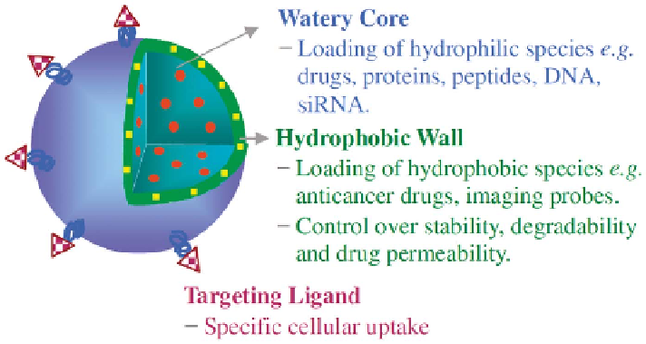
Figure
6.3
Schematic
polymersome
structure,
function,
and
applications.
Zhong
21
(Reproduced
from
Meng
and
with
permission
from
the
American Chemical Society.)
PMPC-PDPA polymersomes for intracellular DOX and DNA release,
26,27
and
Lecommandoux et al. designed pH and temperature dual-responsive poly-
mersomes based on poly[2-(diethylamino)ethyl methacrylate]-PGA (PDEA-
PGA).
28
We designed pH-sensitive degradable polymersomes demonstrating
pH-dependant release of PTX (hydrophobic) and DOX?HCl (hydrophilic)
from block copolymers of PEG and an acid-labile polycarbonate, poly(2,4,6-
trimethoxybenzylidenepentaerythritol carbonate) (PTMBPEC).
29
Battaglia
et al. reported that pH-responsive PMPC-PDPA polymersomes encapsulated
with GFP-encoding DNA plasmid could deliver GFP to primary human
dermal fibroblast cells and Chinese hamster ovary cells.
27
Hubbell et al.
reported oxidation-responsive polymersomes based on PEG-b-poly(propylene
sulfide) (PEG-b-PPS) that underwent rapid destabilization in the presence of
H
2
O
2
(oxidative agent) due to conversion of the PPS hydrophobe into a
hydrophile, poly(propylene sulfoxide) and ultimately poly(propylene sul-
fone).
30
They have also designed reduction-responsive polymersomes based
on a PEG-SS-PPS block copolymer containing an intervening disulfide bond,
wherein cellular uptake and disruption of polymersomes leading to efficient
cytoplasmic release of encapsulated substances were observed in cells following
10 min incubation.
31
There is a high concentration of glutathione tripeptides
(reducing agent) in the cytosol and cell nucleus, which makes reduction-
responsive nano-vehicles highly promising for intracellular drug delivery.
32
Lately, we have designed pH and reduction dual-responsive PEG-SS-PDEA
polymersomes that could encapsulate therapeutic proteins with high efficien-
cies by adjusting the pH to 7.4 and then release the proteins intracellularly
upon cell entry and thus induce greatly enhanced apoptosis of MCF-7 cells
compared to free protein and reduction-insensitive controls (Figure 6.4).
33