Biomedical Engineering Reference
In-Depth Information
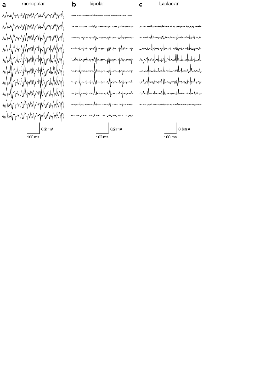
FIGURE 4.56:
Example of spatially and temporally (high-pass at 15 Hz) filtered
signals. Signal from the same 500 ms in monopolar (a), bipolar (b), and Laplacian
(c) derivation. The upper trace corresponds to the medial, the lower to the lateral
electrode positions. From [Kleine et al., 2007].
comparison to signals registered by needle electrode. This problem is now resolved
by application of multielectrode grids, which provide the two-dimensional spatial
information. The differences in location of single MUs with respect to the multielec-
trode and the information about the delay of the given MUAP connected with propa-
gation of MUAPs along the muscle fibers, create so-called MU signatures, which are
utilized in the algorithms of sEMG decomposition.
The semi-automatic techniques for extracting single MUs from sEMG including
operator-computer interaction were proposed in 1999, e.g., [Disselhorst-Klug et al.,
1999]. Later, the fully automatic system for extraction and classification of single
MUAPs from sEMG was designed by Gazzoni et al. [Gazzoni et al., 2004]. The seg-
mentation phase was based on the matched continuous wavelet transform, and was
performed in two steps. In the first step candidate MUAPs were detected by multi-
scale matched filter. The second step was based on the properties of propagation of
the action potentials along muscle fibers. Only MUAPs presenting clear propagation
along the longitudinal fiber direction were selected. The clustering was performed by
a set of neural networks, working in parallel, one for each available channel. They
operated in an adaptive way updating the templates and creating new clusters for
non-matching patterns. However, the system didn't offer the possibility of resolving
the superimposed MUAPs.


Search WWH ::

Custom Search