Biomedical Engineering Reference
In-Depth Information
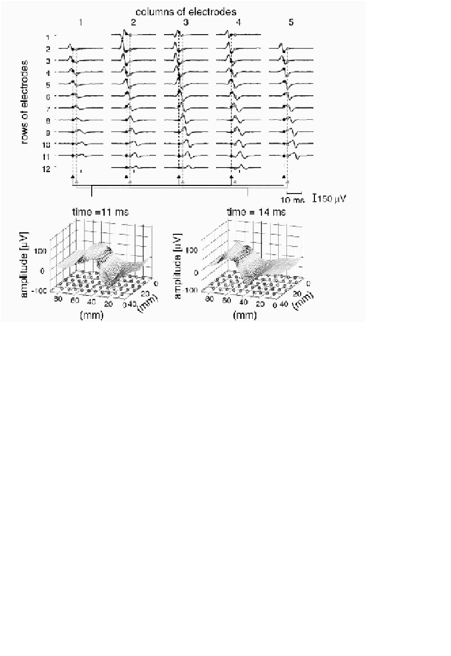
FIGURE 4.54:
Motor unit action potential recorded with a grid of 61 electrodes
(13
5 electrodes, inter-electrode distance 5 mm) from the biceps brachii muscle
during an isometric contraction at 10% of the maximal force. The bipolar signals
derived along the direction of the muscle fibers are shown in the top panel. The elec-
trode grid was placed distal with respect to the innervation zone of the motor unit.
The signals detected along the rows show similar action potential shapes with a delay
corresponding to the propagation along the muscle fibers. The multi-channel action
potential is a three-dimensional signal in time and space. The two-dimensional spa-
tial representations for two time instants (11 ms and 14 ms after the generation of the
action potential) are shown in the bottom panel. The circles on the plane representing
the spatial coordinates mark the locations of the electrodes of the grid. From [Mer-
letti et al., 2008].
×
The biggest challenge in sEMG processing—the extraction of individual
MUAPs—became possible when the high-density grids, including sometimes hun-
dreds of electrodes, became available. Figure 4.54 shows motor unit action potential
recorded with a grid of 13x5 electrodes and illustrates tracking of the propagation
of MUAP by multielectrode arrays. The process of the sEMG decomposition into


Search WWH ::

Custom Search