Biomedical Engineering Reference
In-Depth Information
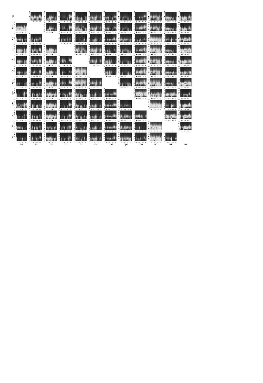
FIGURE 4.21: (SEE COLOR INSERT)
Propagation of EEG activity in the left
hemisphere during right finger movement. In each panel SDTF as a function of time
(horizontal axis) and frequency (vertical axis). The flow of activity is from electrode
marked under the column to the electrode marked at the relevant row. Red—the
highest intensity, blue—the lowest. From [Ginter et al., 2001].
concerning the interactions of brain structures during simple and complicated motor
tasks.
Another application of SDTF concerned evaluation of transmission during cogni-
tive experiments. In the continuous attention test (CAT) geometrical patterns were
displayed and the subject had to react to two subsequent identical pictures by press-
ing the switch (situation target) and do nothing when the patterns were not identi-
cal (situation non-target). The results confirmed the engagement of pre-frontal and
frontal structures in the task and elucidated the mechanisms of active inhibition [Bli-
nowska et al., 2010a].
Figure 4.23
shows the snapshots from the movie illustrating
transmissions. Only significant flows showing increase or decrease of propagation
in respect to reference period are shown. They were calculated according to the
procedure devised in [Korzeniewska et al., 2008], which will be described in
Sect.
4.1.7.3.7.
In Figure 4.23 one can observe for the non-target situation the flow from
electrode F8 (overlying right inferior cortex) to electrode C3. This kind of transmis-
sion was present in six out of nine studied subjects. In three subjects the propagation
for non-target was from the Fz electrode (overlying pre-supplementary motor area).


Search WWH ::

Custom Search