Biomedical Engineering Reference
In-Depth Information
to construct a feature vector capturing the morphology and topography of the EEG
epoch. By means of a support vector machine classifier the vector representative of
seizure and non-seizure epochs was constructed individually for each patient. The
method was validated on 36 subjects. The authors reported 94% sensitivity, and an
average latency in detecting seizure onset of 8s.
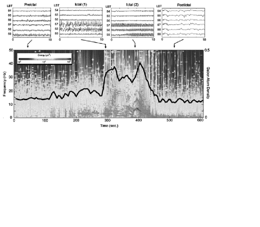
The interesting method of seizure detection based on the decomposition of signal
by means of MP algorithm was proposed by [Jouny et al., 2003]. The method relies
on the observation that at the seizure onset the number of atoms needed to describe
the signal rapidly increases. The authors introduced the measure called GAD (Gabor
atom density). GAD was defined as the number of atoms obtained during the MP
decomposition divided by the size of the reconstructed time-frequency space. The
criterion for termination of iterative decomposition was based on the energy thresh-
old found empirically for all studied subjects. GAD calculated for moving window
together with the time-frequency plot is shown in Figure 4.17. GAD measures com-
plexity of a signal as the number of elementary components needed to represent it.
The method appeared to be very sensitive and specific for detecting intracranial ictal
activity. The earliest changes during a seizure have been observed in the channels
closest to the region of seizure onset.
FIGURE 4.17: (SEE COLOR INSERT)
GAD analysis of a complex partial
seizure. Upper panels: EEG traces for 4 different epochs. Lower panel: color-coded
time-frequency energy density of the signal from contact LBT 58. Superimposed
over this plot is the corresponding GAD (black trace). From [Jouny et al., 2003].


Search WWH ::

Custom Search