Biomedical Engineering Reference
In-Depth Information
time occurrence amplitude, and phase. This allows to construct explicit filters, based
on clinical definition, for finding in EEG specific structures of both transient and os-
cillatory nature. In fact MP is the only method which determines as a parameter the
duration of signal structure, which allows for explicit application of R&K criteria
concerning the percentage of specific activity in the given epoch. One of the first
applications of MP to biological signals concerned sleep spindles [Blinowska and
Durka, 1994]. High accuracy identification and parametrization of sleep spindles by
means of MP allowed for distinction of two classes of spindles of different frequency
ranges and topographic localization [Zygierewicz et al., 1999]. In the following work
the time evolution of SWA and spindle activities was investigated and an inverse re-
lation in their magnitudes was found [Durka et al., 2005a]. Macro and microstructure
of sleep EEG was considered in [Malinowska et al., 2006] and [Malinowska et al.,
2007], where identification of K-complexes, detection of deep sleep stages (3 and
4) based directly upon the classical R&K criteria, continuous description of slow
wave sleep, fully compatible with the R&K criteria, and detection of arousals were
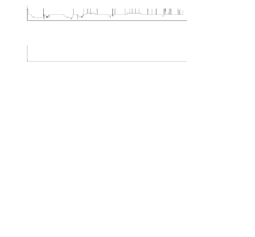
presented in the framework of the same unifying MP approach (Figure 4.13).
S4
S3
S2
REM
S1
W
M
0
1
2
3
4
5
6
7
100
50
20
0
0
1
2
3
4
5
6
7
30
20
10
0
0
1
2
3
4
5
6
7
2
1
0
0
1
2
3
4
5
6
7
400
200
0
0
1
2
3
4
5
6
7
200
100
0
0
1
2
3
4
5
6
7
300
200
100
0
0
1
2
3
4
5
6
7
time [h]
FIGURE 4.13:
Time evolution during overnight sleep of the signal structures
quantified by means of MP. From top to bottom: hypnogram; percentage of 20 s
epoch occupied by waveforms classified as SWA; number of sleep spindles in 3 min-
utes periods; K-complex occurrence; power of theta waves per 20 s; power of alpha
waves per 20 s; power of beta waves per 20 s epoch. By courtesy of U. Malinowska.


Search WWH ::

Custom Search