Biology Reference
In-Depth Information
functional analyses of TH response genes, focusing on potential signaling
pathways involved in establishment of the stem cell niche.
2. ADULT EPITHELIAL DEVELOPMENT DURING
INTESTINAL REMODELING
The larval-to-adult remodeling has been well characterized in the
X. laevis
intestine at the cellular level. Throughout pre- and prometamorphosis,
the long small intestine has a simple tubular structure with only a single lon-
gitudinal fold, the typhlosole, which is localized in the anterior part of the
small intestine (
Marshall & Dixon, 1978b
). Histologically, the intestine
mainly consists of a single layer of the primary (larval) epithelium, the im-
mature connective tissue which is very thin except in the typhlosole, and
thin layers of inner circular and outer longitudinal muscles (
Fig. 11.1
).
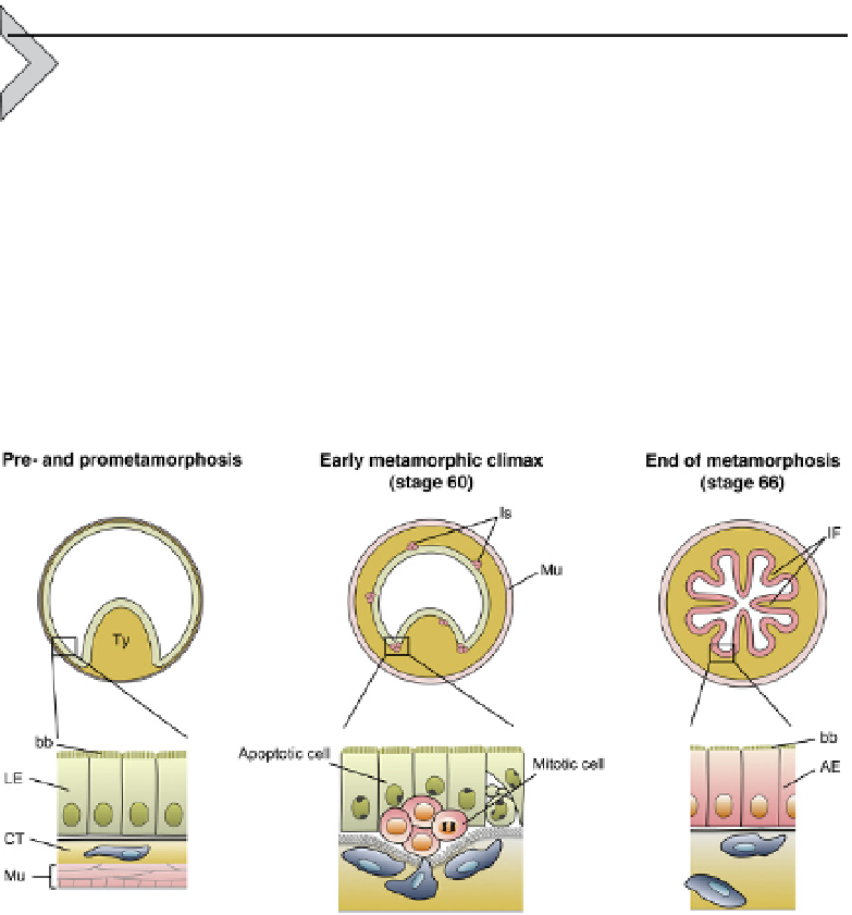
Figure 11.1 Larval-to-adult intestinal remodeling during Xenopus laevis metamorpho-
sis. During pre- and prometamorphosis, the small intestine has a single fold, typhlosole
(Ty), and consists of the larval epithelium (LE) possessing the brush border (bb), the im-
mature connective tissue (CT), and thin layers of inner and outer muscles (Mu). At the
early metamorphic climax (stage 60), most of the larval epithelial cells begin to undergo
apoptosis, whereas adult progenitor/stem cells strongly stained red with pyronin Y ap-
pear as islets (Is) between the degenerating larval epithelium and the developing con-
nective tissue. They actively proliferate and gradually replace the larval epithelial cells.
Then, with the progress of intestinal fold formation, they differentiate into a single layer
of the adult epithelium (AE) possessing the shorter brush border. After the end of meta-
morphosis (stage 66), the adult epithelium acquires a cell renewal system along the
trough
crest axis of the intestinal folds (IF).
-