Biology Reference
In-Depth Information
Degradation
Elongation
and
closure
Docking
and
fusion
Induction
Autophagy
signal
Isolation
membrane
Autophagosome
Lysosome
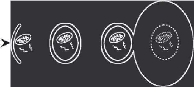
Figure 4.1 Diagram of the steps in autophagy. Autophagy is a catabolic process by
which organelles and cytoplasmic proteins are degraded. Induction of autophagy
results in the formation of an isolation membrane, which expands and closes around
cytoplasmic material, forming the double-membraned autophagosome. The auto-
phagosome traffics to the lysosome where it docks and fuses, releasing its inner mem-
brane and its contents. The autophagosome contents are degraded by lysosomal
enzymes and are recycled back to the cytoplasm through permeases.
important process for maintaining cell homeostasis, responding to stress, and
surviving nutrient starvation.
2.1. Regulatory pathways
Several metabolic regulatory factors affect autophagy induction, including
nutrient availability, insulin signaling, and ATP levels (
Meijer &
Codogno, 2004
). The mechanistic target of rapamycin (TOR) plays a cen-
tral role in autophagy by integrating the class I phosphatidylinositol-3-kinase
(PI3K) and amino acid signaling pathways (
Wullschleger, Loewith, & Hall,
2006
). When nutrients are available, class I PI3K activates TOR, which re-
presses autophagy by phosphorylating Atg13. This hyperphosphorylation
reduces the affinity of Atg13 for Atg1, decreasing the kinase activity of
Atg1 and inhibiting autophagy (
Kamada et al., 2000; Noda & Ohsumi,
1998
). During nutrient starvation, TOR activity is reduced, relieving its re-
pression of Atg1, and autophagy is induced. Increased autophagy contributes
to cell survival by producing amino acids and fatty acids that are used by the
tricarboxylic acid cycle to generate ATP (Lum et al., 2005).
The origin of the autophagic membrane is not completely understood
and remains a subject of debate (
Juhasz & Neufeld, 2006
). In yeast,
autophagy proteins gather at the pre-autophagosomal structure (PAS) near
the vacuole (
Mizushima, 2007
). In animal cells, a PAS-like structure has