Biomedical Engineering Reference
In-Depth Information
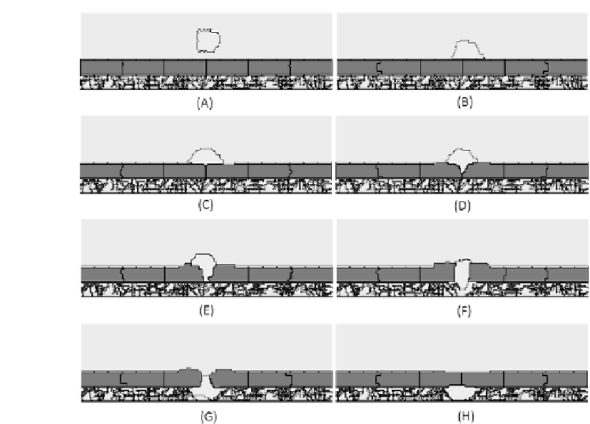
FIGURE 3.6: Simulated single cell transmesothelial migration at 0, 200, 400,
700, 800, 950, 1100, and 1200 MCS. In (B) the cell touches the mesothelial
layer covered by the pericellular matrix and starts spreading over it. In (C) the
production of MMPs starts degrading the pericellular layer. In (D) the cancer
cell induces the loosening of the adhesion bonds between the epithelial cells
that detach so that the cell progressively penetrates the layer. After the cell
has migrated to the opposite side of the layer (G), the simulated mesothelium
has closed back (H), and the tumor cell is considered completely infiltrated.
to set
8
<
T
C
T
M
;
T
E
= 0;
:
surface
C
surface
M
surface
E
;
M
perimete
E
:
The initial hierarchy of the Js is obviously important to maintain the struc-
ture of the mesothelial layer rigid, surrounded by the pericellular matrix and
fixed on the submesothelial ECM fibers. In particular J
M;M
and J
M;E
are
perimeter
C
perimeter


Search WWH ::

Custom Search