Biomedical Engineering Reference
In-Depth Information
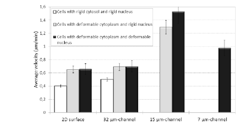
FIGURE 10.5: Comparison of the average migration speed of cells moving
either on the flat surface or within the different microchannels. Cell velocity
on both the bidimensional substrate and in the larger channel is substantially
low, and does not significantly depend on the cytosol and nucleus rigidities.
On the contrary, cell speed within the more confined structures is generally
two-fold higher and is further enhanced by the possibility of nucleus deforma-
bility. The values of the cell average velocity are represented as means s.d.
over 50 \eective" realizations. This means that, for this statistical analysis,
we only consider cells that completely enter within the channel of interest
(i.e., with an invasive or permeative phenotype). The others are classified as
nonmotile, assigned an undefined velocity, and are not taken into account. Fi-
nally, if in a given case the number of cells entering a channel structure is not
significant, we do not evaluate the relative average speed. In particular, sta-
tistical signicance (p < 0.05) is determined via both the Students' t-test for
motile fraction data and via the Kolmogorov{Smirnov test for non-normally
distributed data sets. The specific values regulating the elastic properties of
the cell subcompartments are those used in the relative sets of simulations.
with almost similar values, is observed for cell migration in the largest chan-
nel, confirming that extracellular environments whose dimensions are greater
than cellular measures do not represent guidance cues, but rather behave as
open spaces.
In the case of the intermediate channel, cells with an elastic cytosol display
instead an approximately two-fold increment in cell migration speed ( 1.1
m/min), which is significantly enhanced when allowing nuclear deformability
( 1.5 m/min). The dierent migration speeds of cells within either the big


Search WWH ::

Custom Search