Biomedical Engineering Reference
In-Depth Information
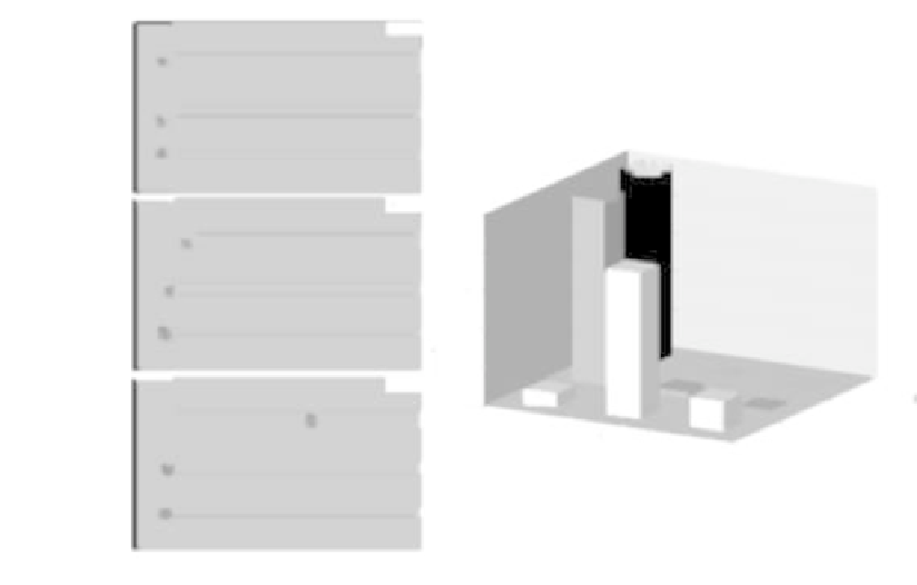
FIGURE 10.2: Migratory behavior within the microchannel structure of can-
cer cells unable to remodel at all (i.e., characterized by a high
surface
;
= 15,
for (
) = fC;Ng). (A) Images of a time-lapse simulation taken at t = 1 (top
panel), 2 (middle panel), and 8 (bottom panel) h. Only the largest channel can
be entered by moving individuals, which, forced to remain in their initial hemi-
spheric morphology, are prohibited from squeezing through smaller structures.
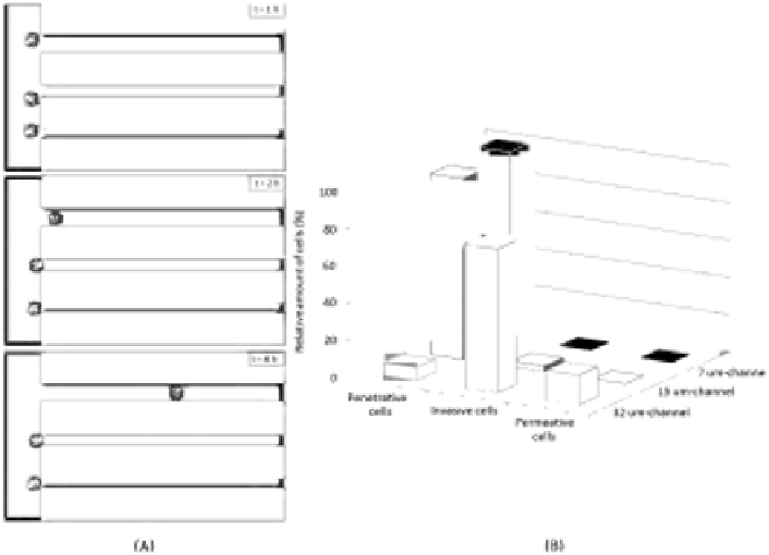
(B) Summary of cell migratory behavior within the matrix device. In the case
of middle and small channels, cells display a penetrative non-invasive pheno-
type, whereas in the case of the biggest channel, they are typically invasive.
The quantitative evaluation of specific cell motile phenotypes, represented in
the histogram plot, is obtained by performing 100 simulations.
counterpart of the activity of phalloidin-like compounds, which block the re-
organization of the cell cytoskeleton by inhibiting actin{myosin interactions.
As reproduced in Figure 10.2, all individuals initially show a random migra-
tion on the flat surface (i.e., until nearly 2 h). Then, when approaching the
microchannels, they start walking along them. In this regard, it is useful to
emphasize that this preferred cell movement is completely autonomous, as we
do not include in the model any chemical gradient or bias, or any a priori
direction for moving individuals.
In the case of the channel with the largest cross{section, the cells typi-
cally display invasive behavior, as they are able to enter and migrate within
the structure, but cannot reach the opposite border, as shown in Figure 10.2.


Search WWH ::

Custom Search