Biomedical Engineering Reference
In-Depth Information
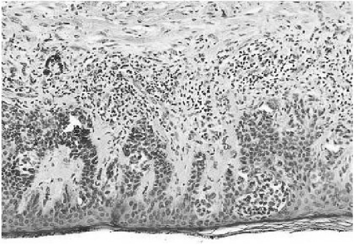
FIGURE 8.10: For comparison purpose, high-power photomicrograph (cour-
tesy of I.R.C.C.) of a lentiginous and junctional moderate melanocytic dys-
plasia in the epidermis overlying the dermal component. The papillary dermis
is widened by a mixture of tumor cells, inflammatory cells, and fibrous tissue.
At the bottom of the image, invading fronts of the neoplasm feature tentacu-
lar or finger-like extensions, similar to those reproduce by the computational
model.
full-size cells. We further assume that both daughter cells evenly inherit all the
parent's biophysical properties (i.e., its motility and adhesive properties). Fi-
nally, the newly formed individuals are placed symmetrically about the parent
cell center of mass with a random orientation.
Intuitively, cell proliferation is expected to be dramatically proinvasive, on
the basis that an increment in the cell population will facilitate the invasion
of the extracellular environment. However, tumor invasiveness only slightly
increases, as df
f
260 m (see Figure 8.9). The explanation of this counter-
intuitive result is that the external cells, whose metabolism is accelerated
by the high quantity of available nutrients, quickly divide, forming a front
of little islands, as reproduced in Figure 8.9(B). Such cell clusters go on in-
creasing in size, due to further cell proliferations, and come in contact with
the main tumor mass by short and thick (4{5 cell-wide) \ngers" (see Fig-
ure 8.9(C)). The increased cellular density, in turn, enforces cell{cell adhesive
interactions, which balance the effect of the haptotaxis and of the enhanced
cell motility. The formed fingers therefore do not break and, consequently, the


Search WWH ::

Custom Search