Biomedical Engineering Reference
In-Depth Information
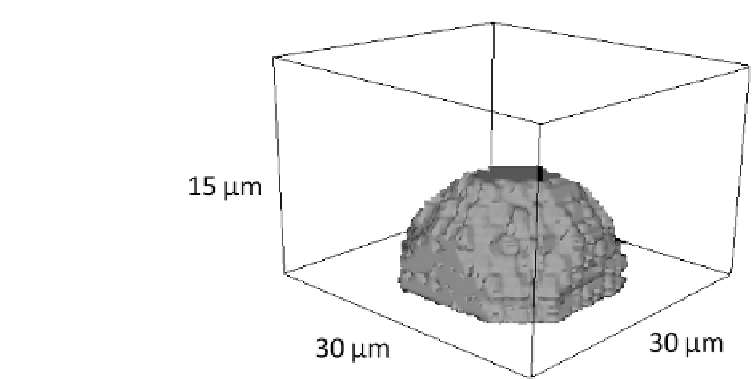
FIGURE 6.2: Representation of the initial 3D morphology of the simulated
tumor-derived EC, which is 30 m in length and width and about 15 m in
height.
C
0
=
P
x
2
c
0
is its total basal level, i.e., the level below which the cell dies.
sur
0
is instead the intrinsic cell resistance to compression at the basal cal-
cium amount C
0
: observing that resting TEC maintain their initial geometrical
configuration, with negligible changes of shape or cytoskeletal active reorga-
nization, we have chosen a high value for
surf
0
. In particular, for saturating
and for
such that (
) = C,
surface
levels of Ca
2+
;
! 0, and the cell
can undergo dramatic changes in its morphology in response to the external
stimulus.
H
adhesion
takes into account only the generalized adhesion between the
nucleus and the cytosolic compartments, which, as seen, is a general extension
of the Steimberg's Dierential Adhesion Hypothesis (DAH), refer to Equation
(4.2). To prevent the cell to split into disconnected patches, we assign a large
negative energy penalty J
int
C;N
.
Since vascular ECs have been demonstrated to migrate along gradients
of VEGF concentration [157, 360], we add a classical linear-type chemotaxis
term of the form (1.9):
H
chemotaxis
=
ch
;
(
x
source
)
(x
source
;t) [q(x
target
;t) q(x
source
;t)] ; (6.2)
where x
source
and x
target
are, as usual, the source and the final lattice sites
randomly selected during a trial update in a MCS and (
(
x
source
)
) = C.
Obviously, x
source
is a site belonging to the border of the cytosolic region,
while x
target
is a medium site. The parameter
ch
;
(
x
)
represents the local
chemical sensitivity of the cell and evolves according to a Michaelis{Menten


Search WWH ::

Custom Search