Agriculture Reference
In-Depth Information
A
perioplic groove (1)
coronary groove (2)
epidermal lamellae (3)
bulb of the heel (7)
hoof wall (4)
bulb of the hoof (6)
B
C
2
1
3
sole (5)
7
7
quarter
toe
6
5
4
6
perioplic corium (8)
E
G
D
coronary corium (9)
dermal
lamellae (10)
6
10
white zone
5
solear corium
8
F
4
9
10
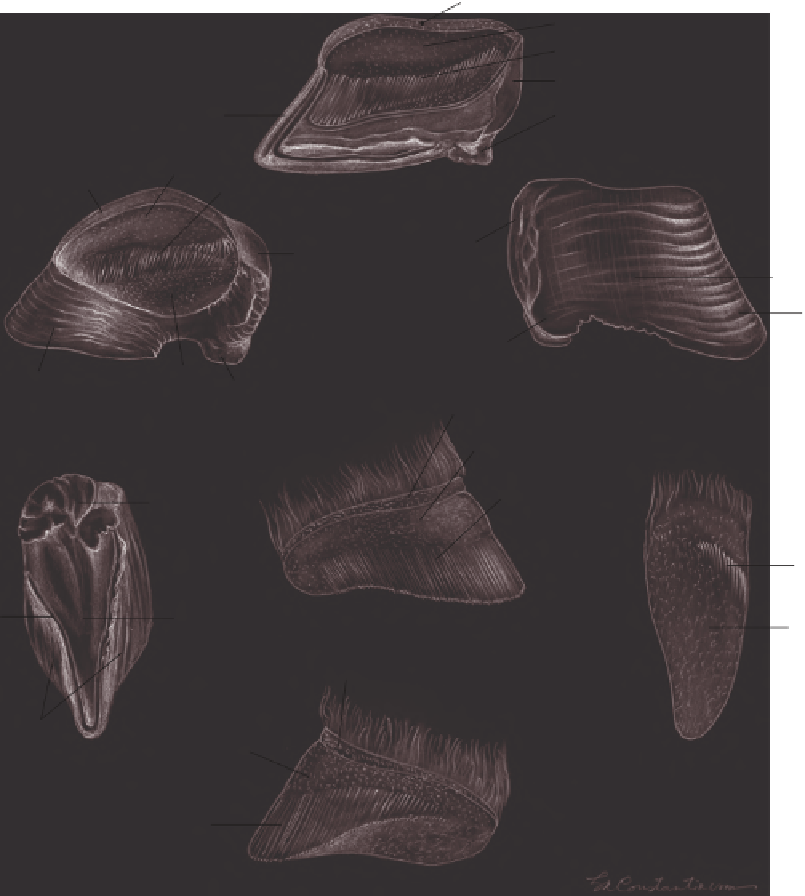
Figure 6.20
The hoof and the dermal coria: A. Median section; B. Inner aspect of the hoof; C. The
hoof wall; D. The solear aspect of the hoof; E. Dermal corium, abaxial aspect; F. Dermal corium, axial
aspect; G. Dermal corium, solear aspect.
The inner surface of the hoof wall corresponds to the
structures of the parietal corium, which are protected by
the wall. In a proximodistal direction, they are the perioplic
groove, the coronary groove, and the horny lamellae
(laminae). The latter are extremely numerous (between
550 and 700). The horny lamellae are white in color
regardless of hoof wall color, and are parallel with each
other. They end in small papillae and will show up on the
solear aspect of the hoof between the sole and the wall of
the hoof as the “white zone” (see Figure 6.20). The digital
arteries and veins and the digital nerves supply heavily the
live tissues of the hooves.
THE CARDIOVASCULAR SYSTEM
The cardiovascular system consists of the heart, the blood
vessels (arteries, veins, and capillaries), and the lymphatic
system (lymph nodes and lymph vessels). The spleen is
associated with the cardiovascular system, but it will be