Biology Reference
In-Depth Information
Retinal ganglion
Cells (melanopsin)
RHT
SCN
POT
SPVZ
IGL
GHT
PTA
RPT
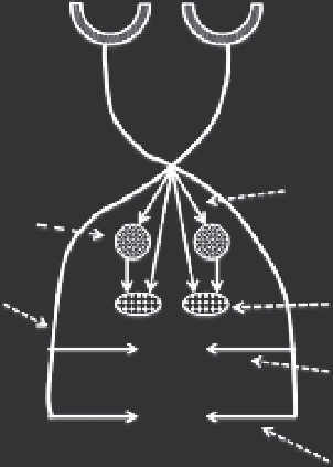
Figure 1.6 Diagram showing retinal projections to the components of the circadian
timing system. IGL, intergeniculate leaflet; GHT, geniculohypothalamic tract; PTA,
pretectal area; POT, primary optic tract; RHT, retinohypothalamic tract; RPT, ret-
inopretectal tract; SCN, suprachiasmatic nucleus; SPVZ, subparaventricular zone.
4.3. Efferent organization
distinguishable from other systems. Early studies used an autoradiographic
ing of fibers of passage from terminal plexuses. The most extensive data
come from studies on the rat using anterograde transport using
Phaseolus
leucoagglutinin (Refs.
117-119,150
)
. These showed a pattern of efferents
distributing predominantly to adjacent hypothalamus, preoptic area,
anterior hypothalamic area, particularly the subparavenricular zone, retro-
chiasmatic area, tuberal and posterior hypothalamic areas, as well as more
limited projections to basal forebrain, midline thalamus, IGL, and
projections to these areas arise differentially from core and shell (Refs.
prokineticin 2 (PKC2), a member of a family of secreted proteins with
protein is found in SCN neurons and projections and appears necessary
for transmission of the circadian signal (Refs.
121,122
).

Search WWH ::

Custom Search