Information Technology Reference
In-Depth Information
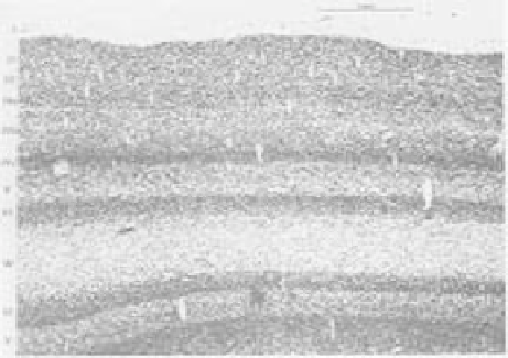
Figure 3.2:
Image of a slice through the visual cortex of a
cat, with the neuron cell bodies stained, showing the six pri-
mary layers, plus the different sublayers of the input layer 4.
Reproduced from Sejnowski and Churchland (1989).
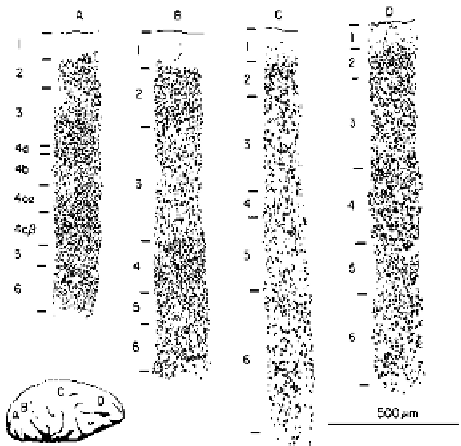
Figure 3.4:
Different laminar structure of cortex from differ-
ent areas. A) shows specialization for the input layer 4 in the
primary visual input area. B) shows emphasis on hidden lay-
ers 2/3 in a hidden area (extrastriate cortex) higher up in the
visual processing stream. C) shows emphasis on output layers
5/6 in a motor output area. D) shows a relatively even blend
of layers in a prefrontal area.
Reproduced from
Shepherd
Hidden
(Layers 2,3)
(1990).
Input
(Layer 4)
Output
(Layers 5,6)
much remains to be done.
The view of the cortex presented in figure 3.3 has
been simplified to emphasize the distinctive laminar
(layered) structure of the cortex. In reality, the cortical
areas that process sensory input are not the same ones
that produce motor output, and there are a large num-
ber of areas which have neither direct sensory input nor
direct motor output. Thus, figure 3.5 presents a more
accurate (though still abstracted) picture of the struc-
ture of the three main different kinds of cortical areas,
which correspond to the three functional layer types (
in-
put, hidden,
and
output
). Each of these different types
of areas emphasizes the corresponding functional layers
(figure 3.4) — input areas have a well developed corti-
cal layer 4 (including different sublayers of layer 4 in
the primary visual input area of the monkey), and out-
put areas have well developed output layers. The hidden
Sensation
(Thalamus)
Subcortex
Motor/BG
Figure 3.3:
A simple, three-layer interpretation of cortical
structure that is consistent with general connectivity patterns
and provides a useful starting point for modeling. Direct ex-
citatory connectivity is shown by the open triangular connec-
tions. Inhibitory interneurons are indicated by the filled circu-
lar connections; these operate within each cortical layer and
receive the same types of excitatory connections as the exci-
tatory neurons do. Dotted lines indicate connections that may
exist but are not consistent with the flow of information from
input to hidden to output. Limited data make it difficult to
determine how prevalent and important these connections are.


Search WWH ::

Custom Search