Information Technology Reference
In-Depth Information
V2 neurons exhibit their feature selectivity
across a
range of different positions
. Thus, these neurons can be
seen as the initial steps toward spatially invariant object
representations. We will see in section 8.4 how these
kinds of partially invariant V2 receptive fields might de-
velop, and how the weights need to be configured to
achieve this property.
The next major area after V2 is
V4
, which receives
inputs from V2, and is the first visual area that appears
to be primarily focused on visual form processing and
object recognition. Here, neurons continue the process
of spatial invariance coding begun in V2, and also ex-
hibit more complex feature detection.
In the inferior temporal cortex (
IT
), neurons achieve
a high level of both size and location invariance, and
some measure of rotational invariance. Further, the neu-
rons encode complex (and often very difficult to char-
acterize) properties of shapes. Thus, it seems likely that
IT neurons provide a distributed basis for invariant ob-
ject recognition. Lesions in this area can produce
visual
agnosias
, or the inability to recognize objects visually
(Farah, 1990). One particularly interesting form of ag-
nosia is
prosopagnosia
, where a lesion results in the in-
ability to recognize faces (see Farah et al., 1993 for a
neural network model). For more details on some of
the specific properties of this neural code, see
m
m
PO
VIP
PG
V3A
MST
FST
p
MT
PG
V1
V2
V1
d
TE
V3
V4
TEO
TE
TF
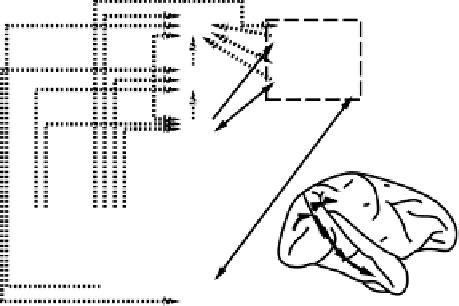
Figure 8.6:
Diagram of the monkey visual system, showing
the dorsal “where” pathway (to PG) and the ventral “what”
pathway to TE. Solid lines indicate full visual field projec-
tions, whereas dotted lines are only the peripheral visual field.
Adapted from Desimone & Ungerleider (1989), which con-
tains further details.
to be functionally isolated processing “modules.” We
will explore one idea about what these interconnections
might be doing in the models described later in this
chapter.
Tanaka
(1996) and Desimone and Ungerleider (1989).
8.2.6
The Dorsal Where/Action Pathway
8.2.5
The Ventral Visual Form Pathway: V2, V4,
and IT
The dorsal pathway represents spatial information and
other information relevant for action. Neurons in this
pathway have large receptive fields, often have pre-
ferred directions of motion, and incorporate information
about the position of an animal's head and eyes. All of
these properties support the processing of information
about the location of objects in this pathway.
The dorsal pathway areas proceed up to the parietal
lobe, and they include areas such as
MT
and
MST
(important for motion processing) and posterior pari-
etal areas such as
VIP
and
LIP
. There is considerable
evidence that these areas process spatial information
(e.g., Andersen, Essick, & Siegel, 1985; Ungerleider &
Mishkin, 1982). Perhaps the most dramatic evidence
comes from patients with
hemispatial neglect
.These
The ventral pathway for representing visual form infor-
mation (i.e., object recognition) can be thought of as
successive stages that lead to increasingly spatially in-
variant representations. In addition, the complexity of
the form information encoded by these representations
increases in successive stages, and the receptive fields
get larger.
ThenextareaafterV1,called
V2
, appears to con-
tain a number of interdigitated regions (called
stripes
)
of specialized neurons that emphasize different aspects
of visual information (e.g., form (edges), surface prop-
erties (color, texture), and motion). As emphasized by
Desimone and Ungerleider (1989), one critical differ-
ence between V2 and V1 representations is that some


Search WWH ::

Custom Search