Information Technology Reference
In-Depth Information
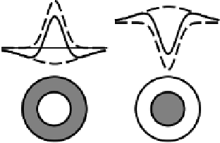
a) On−center
b) Off−center
tribution (in the retina) of inputs (light) that affect the
firing of that neuron (more generally, this term refers to
the set of inputs that activate a given neuron). You can
picture the center-surround RF as a target, with a cen-
tral region and a surrounding ring. Figure 8.2 shows the
two main categories of center-surround receptive fields:
an
on-center
RF, where the neuron is more active when
the central portion of its receptive field is brighter than
the surrounding portion, and an
off-center
RF, where
the neuron is more active when the center is darker than
the surround.
As illustrated in the figure, the on-center RF is con-
structed by subtracting a broadly-tuned but somewhat
shallow surround region from a more sharply tuned
and more peaked center region, and vice-versa for
the off-center RF. Because each of the individual tun-
ing functions can be modeled by a Gaussian (normal,
bell-shaped) distribution function, the resulting center-
surround field has been called a
difference of Gaus-
sians
(DOG).
Consider what would happen if a uniform region of
light covered the entire receptive field of either an on-
or off-center neuron — the excitation would cancel out
with the inhibition, leaving no net effect. However, if
light were to fall more in the excitatory center of an
on-center neuron than in its inhibitory surround, there
would be net excitation. Conversely, if there was
less
light on the inhibitory center of an off-center neuron
compared to its excitatory surround, it would become
excited. These receptive field properties lead to the
compression effect mentioned above, because the reti-
nal output neurons (ganglion cells) fire maximally when
there is a change in illumination level over their recep-
tive fields, not when there is constant illumination. Al-
though we have focused only on intensity (brightness)
coding, retinal neurons also have on- and off-center
coding of different wavelength tunings based on inputs
from the different cone types (i.e., for color perception).
We will see in subsequent sections how these basic
retinal receptive field properties provide useful building
blocks for subsequent processing areas.
+
+
+
+
+
−
−
−
−
−
−
+
+
−
−
+
+
−
+
−
Figure 8.2:
On- and off-center receptive fields computed by
the retina. The bottom shows a two-dimensional picture of
the receptive field, with central and surround regions. The
upper profiles show a slice through the center of the two-
dimensional receptive fields, showing how a broad surround
field and a narrow central field (in dashed lines) can be com-
bined to form the overall receptive field (in solid line) as the
difference of the two fields modeled as Gaussians.
sensitive
photoreceptor
cells that turn photons into
electrical signals. There are two types of photorecep-
tors,
rods
and
cones
. Rods are primarily responsible for
vision under low light levels (
scotopic
vision). Cones
have three different wavelength sensitivities, and are
primarily responsible for vision with high levels of light
(
photopic
vision). Cones are primarily found in the
fovea
, the central part of the retina, which processes the
portion of the image one is directly looking at.
Subsequent stages of retinal processing combine
these electrical signals across local regions of photore-
ceptors, and then perform various types of
subtractions
of different regions using specialized, retina-specific
mechanisms. These subtractions provide the crucial
contrast enhancement effect, and are also responsible
for enhancing the wavelength-selective properties of the
photoreceptors to enable the perception of color. Fi-
nally, the
ganglion
neurons provide the output signals
from the retina to the thalamus, described in the next
section. The retina represents the central region of vi-
sual space, called the
fovea
, with the highest level of
resolution for a highly detailed picture of things in the
focus of one's gaze.
Most of the subtractions computed by the retina in-
volve a
center-surround
receptive field. The
receptive
field
(RF) of a neuron generally refers to the spatial dis-
Search WWH ::

Custom Search