Biomedical Engineering Reference
In-Depth Information
rate increase. Thus, the risk of Wenckebach behavior occur-
ring is minimized in cases in which the atrial rate exceeds the
MTR, the occurrence of great changes at the upper limit is
minimized, and 1:1 tracking at higher rates is enabled.
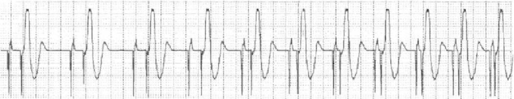
Figure
9.10
shows an ECG record with dynamic AV delay.
When the AA atrial interval is between the LRL and the
higher of the MTR or maximum sensor rate (MSR) values,
the pacemaker determines the dynamic AV delay according
to the preceding AA interval based on linear dependence, as
shown in Fig.
9.11
. This relation is determined by pro-
grammed values of minimum AV delay, maximum AV delay,
LRL, and the higher of the MRT or MSR values.
Another possibility of compensating for the time differ-
ence between a paced and spontaneous atrial event is the
activation of the SAV offset function. As a result, the AV
delay is shortened after a sensed atrial event by a programmed
value. As a consequence, the hemodynamic AVI is different
for paced and sensed atrial events. If a constant AV delay is
set, the SAV offset parameter will also be fixed at a pro-
grammed value. If a dynamic AV delay is set, the pacemaker
calculates the SAV offset parameter based on the intrinsic
atrial rate. As a response to narrowing of the P wave in the
period of increased metabolic requirements, the SAV offset
is shortened linearly from a programmed value correspond-
ing to the LRL to a value determined by the proportion of
minimum AV delay and maximum AV delay and the higher
of the MRT or MSR values.
9.6
Tracking Atrial Rhythm to Ventricles
In tracking modes of dual-chamber pacing, sensing and pac-
ing in either the atrium or ventricle may occur, or in the case
of biventricular pacing, possibly in both ventricles. In a
tracking mode, the device responds to a sensed intrinsic atrial
event by planning ventricular pacing. The delay between a
sensed atrial event and corresponding ventricular pacing rep-
resents a programmed SAV. If an existing AEI is not termi-
nated quickly by a sensed intrinsic atrial event, the device
paces the atrium and then plans ventricular pacing to be
delivered after a programmed PAV. If a ventricular event is
sensed during the SAV or PAV, ventricular pacing is inhib-
ited. A sensed atrial event occurring during the PVARP is
classified as refractory; it does not inhibit atrial pacing and is
not tracked. DDD(R) is the only mode to be considered a
fully dual-chamber and tracking pacing mode. In the absence
of intrinsic atrial activity, pacing in the DDD mode is deliv-
ered at a programmed LRL. In DDD(R) mode with adaptive
pacing rate, pacing is delivered at a sensor-indicated rate.
9.6.1
Dynamic AV Delay
Shortening the AV delay with an accelerated cardiac action is
a physiological response of the heart. The AVI may be either
programmed to a fixed value or calculated dynamically based
on the preceding AA interval. With a constant AVI, it is difficult
to set an optimum value of the AVI to meet the patient's needs.
At higher rates, a short AVI is appropriate to avoid symptom-
atic 2:1 block during loading and asynchronous pacing.
At lower rates, a long AVI is appropriate to support the intrin-
sic AV conduction; hemodynamics may thus improve. So, if a
constant AV delay is programmed, the AV delay value remains
unchanged upon the increase of the heart rate. When using a
dynamic AV delay, more physiological AV coupling is
achieved in the entire range of programmed rates; the size of
the sensing window is maximized at higher rates by automatic
shortening of a PAV or SAV after each interval upon the atrial
9.6.2
Upper Rate Behavior
Because of the risk of induction of ventricular tachycardia by
fast ventricular pacing, a dual-chamber pacemaker working
in the DDD(R) mode may safely track the atrial rhythm only
up to a certain rate. The upper rate behavior occurs if the
patient's intrinsic atrial rhythm is faster than the MTR,
exceeds the limits of atrial sensing determined by TARP, or
both. It occurs only in patients with AV conduction failure;
with normal AV conduction, the intrinsic atrial action is
spontaneously conducted to ventricles.
Fig. 9.10
Dynamic atrioventricular delay at a surface electrocardiogram [32] (© 2012 Boston Scientific Corporation or its affiliates. All rights
reserved. Used with permission of Boston Scienti fi c Corporation)