Biomedical Engineering Reference
In-Depth Information
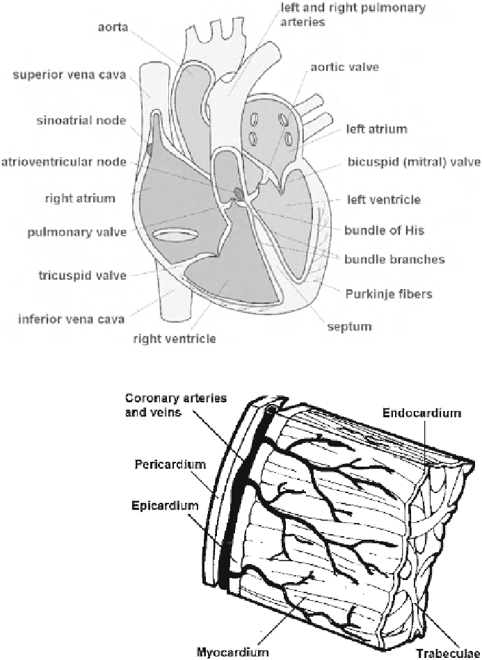
Fig. 3.1
Heart cross
section and conduction
system
3.2
Structure of the Heart Wall
The wall of the heart is composed of three layers: the epicar-
dium, the myocardium, and the endocardium (Fig.
3.2
). The
epicardium is a serous outer sac of the heart wall overlying a
thin layer of elastic fibrous tissue, which attaches the epicar-
dium to the myocardium. Here, fatty tissue is found in places,
particularly in the depressions along the superficial cardiac
arteries, veins, and nerves.
The myocardium is a muscular layer made up of a special
type of striated heart muscle. It is the main component of the
heart wall and has the greatest thickness. It consists of fibers
and individual cells connected in a spatial network. Myocardial
cells have an oval nucleus surrounded by contractile myo fi brils
that have a structure similar to that of skeletal muscle fibers.
The myocardial layer in the atrial walls and septum is much
thinner than that in the ventricles. There is more connective
tissue between muscle stripes in the atrial myocardium than
in the ventricles. The muscle of the left ventricular wall and
septum is about three times as thick as that of the right ven-
tricle. The atrial myocardium is composed of two layers: deep
and superficial. Arches and rings of the deep layer encircle
each of the atria separately. The superficial layer forms longer
transverse stripes that pass from one atrium to the other. The
ventricular myocardium has three layers that are mutually
intertwined to form a common system.
The endocardium is an intracardial membrane that lines
the cardiac cavity. It is smooth, transparent, and glistening. It
consists of a single layer of flat endothelial cells overlying
connective tissue with collagen and elastic fibers. Elastic
Fig. 3.2
Cardiac walls and coverings
fibers are more abundant in the atria than in the ventricles.
There are smooth muscle cells in the connective tissue layer;
these are more abundant in the atrial and ventricular septa.
Endocardial thickness is 50-200 mm. Where thicker, the
endocardium is a whitish color; where thinner, the myocar-
dial muscle is visible.
3.3
The Conduction System
The conduction system is a network of specialized cellular
structures that generate and conduct impulses. The action of
the myocardium is not dependent on additional innervation,