Biomedical Engineering Reference
In-Depth Information
the chromatographic separation. The diff erent phospholipids were
quantified using low-phosphorus silica gel thin layer plates. The
reduction in PG plasma concentration with time was used to calculate
liposome plasma clearance, while the change of ratio of DOX to PG in
the plasma was used to calculate rate of DOX release from DOX-OLV
in the patients' plasma.
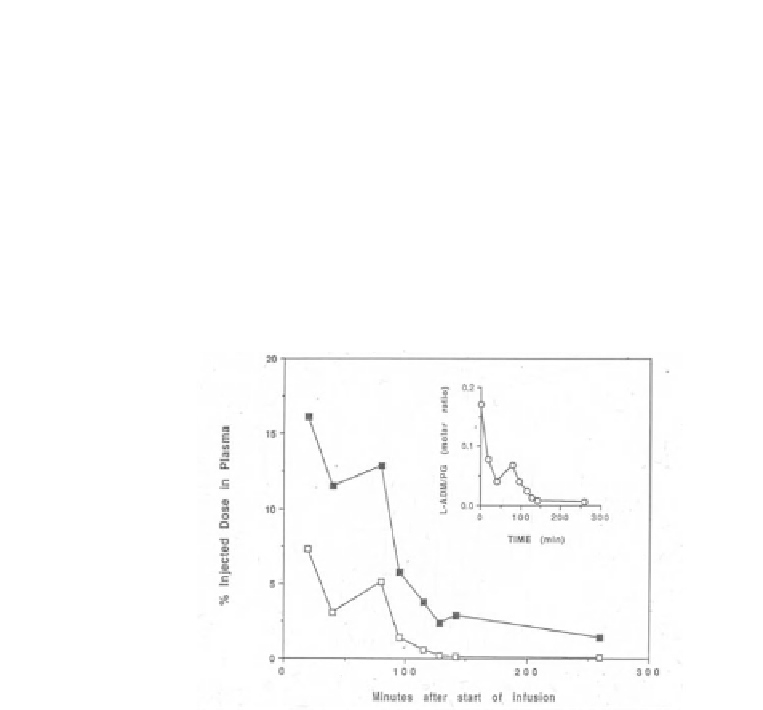
The results obtained in these three patients indicate that
liposome-associated doxorubicin has fast clearance, which resembles
that of F-DOX. In addition, their ratio of DOX/PG is reduced fast
(Figure 12.3, inset) suggesting that liposomes lose most of their
drug payload during circulation.
Figure 12.3
Plasma clearance of liposomal doxorubicin (white squares) and
PG (black squares). Due to dilution-induced release in plasma, the liposomes
reached the liver almost drug free. Adriamycin (ADM) = doxorubicin (DOX).
(Gabizon et al., 1991a; Amselem et al., 1993a)
To learn about the liposome organ distribution, imaging studies
were carried out with doxorubicin-free
111
indium-deferoxamine-
labeled liposomes of the same composition and size distribution as the
OLV-DOX. These
111
In-OLV were prepared by hydration of the lipids
in a medium containing 200 mM of the chelating agent deferoxamine
mesylate (DF) dissolved in physiological saline. Un-encapsulated
DF was removed by passage through a Dowex-50 cation-exchange
resin. Liposomes were labeled with
111
In by incubation with an
111
In-oxine (Amersham) complex at room temperature for about
30 min. Free (unloaded
111
In-oxine was removed by gel permeation


Search WWH ::

Custom Search