Biomedical Engineering Reference
In-Depth Information
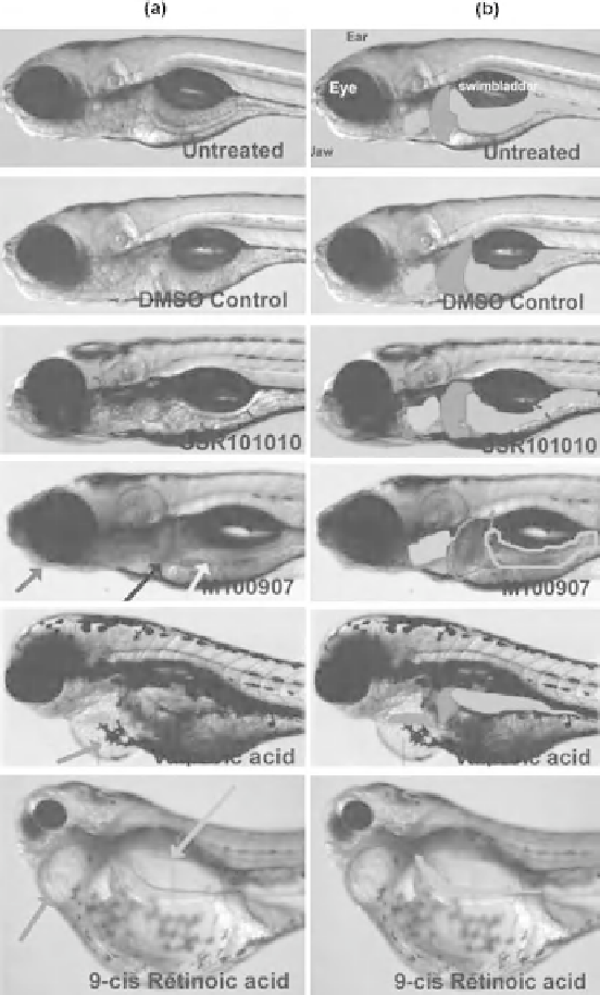
Figure 2.2
Organ structure of treated zebrafish at high magnification (8). Panels (a) and (b) are
the same images; however, organs are outlined in panel (b) to highlight dysmorphogenesis: heart (blue),
liver (red), and intestine (green). SSR101010-treated zebrafish exhibited similar morphology as controls
in all tested concentrations, although precipitation was observed at concentrations 5 mM. M100907-
treated zebrafish exhibited abnormal jaw (blue arrow (a)), and dark opaque liver (black arrow, (a)) and
intestine (yellow arrow, (a)), although organ morphology was not affected (outlined in (b)). In addition,
arrhythmia was observed (data not shown). Valproic acid-treated zebrafish exhibited pericardial edema
(red arrow in (a)); therefore, the heart chambers were stretched to form a thin tube (b). In addition,
small liver and intestine were observed (b). 9-cis-Retinoic acid-treated zebrafish exhibited pericardial
edema and a stretched tiny heart chamber (b); small intestine and absence of liver tissue were also
observed (b). (See the color version of this figure in Color Plates section.)
Search WWH ::

Custom Search