Biomedical Engineering Reference
In-Depth Information
(a)
(b)
Glass capillary
(1.2 mm)
Spacer
(0.35 mm)
Zebrafish
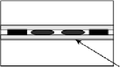
Figure 14.1
Specially designed slide facilitated dye injection into zebrafish brain.
(a) Cartoon illustrating slide design for retaining zebrafish in dorsal orientation to facilitate brain
injection and image analysis. Three rows can be included on each slide and five zebrafish can be
maintained in each row. (b) Image of 4dpf zebrafish, dorsal orientation.
14.2.5 Specialized Zebrafish Slide for Brain Injection
and Imaging
Since maintaining individual zebrafish in a dorsal orientation to facilitate brain
injection and image capture using a conventional depression slide was difficult, we
designed a specialized zebrafish slide that increased throughput. As shown in
Fig. 14.1a, rows of glass capillaries, separated by 0.35 mm spacers, were attached
to slides. Zebrafish were placed between capillaries in the dorsal position, five
zebrafish per row, and at least three rows can be placed on each slide. Using this
specialized slide, the number of zebrafish injected and imaged in 1 h increased from
6to30.
14.2.6 Brain Dye Injection
Zebrafish were placed on slides with 0.32 mM tricaine mixed with appropriate
solution for each condition to immobilize animals. Using a stereo dissecting
microscope (Stemi 2000, Zeiss) for visualization, an injection needle containing
fluorescent rho-HRP (rhodamine-labeled horseradish peroxidase) Pgp substrate
(50mM) (21st Century Biochemicals, Marlboro, MA) was inserted into the midline
of the optic tecta bordering the cerebellum, and 30 nL of substratewas delivered using
a pressure-controlled microinjector (PV-830 Pneumatic PicoPump, WPI) (Meng
et al., 2004) (Fig. 14.2).
14.2.7 Quantitative Morphometric Analysis
of Brain Images
Dorsal view images of brain ROI (region of interest) (25
) were captured using the
same exposure time and fluorescence gain. Fluorescence was quantified using ImageJ
software. A constant threshold was applied to fluorescent brain images of each
zebrafish. This threshold was automatically set by the software and adjustedmanually
for artifacts, when necessary. Total fluorescence from threshold-processed images


Search WWH ::

Custom Search