Biomedical Engineering Reference
In-Depth Information
zebrafish (Parng et al., 2007), supporting use of zebrafish as a model organism for
assessing demyelination and probably remyelination.
10.3.4 Compound-Induced Brain-Specific Apoptosis
Although apoptosis occurs naturally during development of the nervous system,
increased apoptosis in the mature nervous system is deleterious and a hallmark of
many neurodegenerative diseases. Apoptosis is also a common cellular response to
exposure to chemical toxins (Corcoran et al., 1994). Apoptosis is induced in a number
of mammalian cell lines, including neuronal cell lines (Ahmadi et al., 2003; Caughlan
et al., 2004) and mouse embryos after exposure to pesticides (Greenlee et al., 2004).
Organophosphate pesticides induce apoptosis in cultured rat cortical cells (Kim
et al., 2004). Conventional assay formats include absorbance, fluorescence, lumi-
nescence for
in vitro
detection of caspase enzymatic activity, intracellular oxidation,
mitochondrial permeability, or double stranded DNA breaks. However, these
in vitro
approaches have a common shortfall: none of the assays can predict if compounds will
work under
in vivo
physiological conditions.
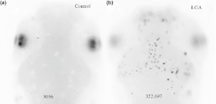
In recent investigations, using both fluorescent acridine orange (AO) live staining
(Parng et al., 2004; Ton et al., 2006) and terminal deoxynucleotidyl transferase dUTP
nick end labeling (TUNEL), zebrafish treated with
L
-2-hydroxylglutaric acid (LGA)
exhibited brain-specific apoptosis (Fig. 10.4) (Serbedzija et al., 2001; Parng et al., 2006;
McGrath et al., 2010). Accumulation of LGA in cerebrospinal and other body fluids has
been observed in patients with hydroxyglutaric aciduria (OHGA) (Hoffmann
et al., 1993). In addition, LGA has been shown to promote oxidative stress and
Figure 10.4
LGA-induced brain apoptosis. After TUNEL staining, images were captured, inverted,
and used for morphometric analysis. Apoptotic cells display a punctate staining pattern in the brain.
Fluorescent signals from the brain region are quantified using ImageJ software (NIH). Total
fluorescence was calculated as fluorescence intensity
staining area. Compared to control (a), LGA-
treated zebrafish (b) exhibited increased apoptosis (3056 versus 322,697).
Search WWH ::

Custom Search