Biomedical Engineering Reference
In-Depth Information
7.3.2 Adult Blood Cell Morphology
There are several ways to assay blood cells in the adult zebrafish kidney. One
possibility is to section the fish, stain with hematoxylin and eosin, and look at
general kidney morphology. In a normal kidney, the hematopoietic cells will be
distributed among the renal tubules (Fig. 7.2a). If the fish has developed a
myeloproliferative disorder, the hematopoietic cells will have taken over the kidney
and fewer tubules will be apparent (Le et al., 2007). Alternatively, if the fish has been
irradiated, the kidney will have few hematopoietic cells when compared to healthy
fish (Traver et al., 2004). Analysis of kidney sections can provide information about
gross hematopoietic development, but does not provide specific information about
blood cell number, blood cell types present in the hematopoietic system, or
individual cell morphology. To look at individual cell morphology and percentages
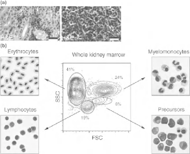
Figure 7.2
Zebrafish kidney marrow. (a) Hematoxylin and eosin staining of zebrafish kidney marrow
sections (Le et al., 2007). A healthy kidney (left) comprised of hematopoietic cells scattered in between the
renal tubules. A fish with myeloproliferative disorder (right) has blood cells overtaking the kidney marrow.
(b) Flow cytometry analysis of zebrafish whole kidney marrow (Traver et al., 2003b). Four populations can
be separated after flow cytometry sorting by forward scatter and side scatter—erythrocytes, lymphocytes,
myelomonocytes, and precursors. Erythrocytes are present in a low FSC population. Lymphocytes are
found in a low SSC and intermediate FSC population. Myelomonocytes fall into a high FSC and SSC
population. Finally, precursors of these cell types have high FSC and intermediate SSC properties. (See the
color version of this figure in Color Plates section.)
Search WWH ::

Custom Search