Biomedical Engineering Reference
In-Depth Information
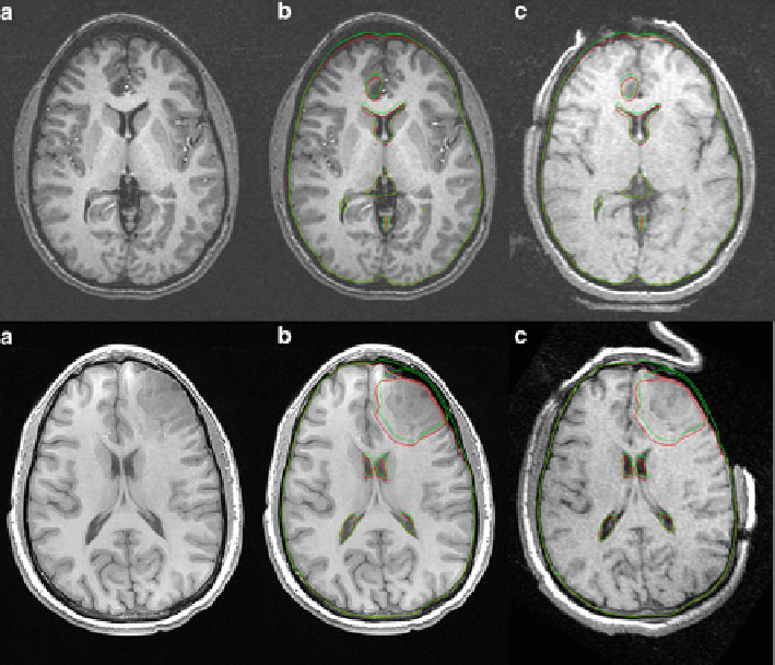
Fig. 2 Registration of 3D MRI images using the deformation field from a biomechanical model.
Two cases are presented: case 1 on the first row and case 2 on the second row. Only one slice is
shown for each case. (a) Preoperative image. (b) Registered pre-operative image. (c) Intra-
operative image (for comparison). Contours of the un-deformed and deformed mesh (including
brain tissue, tumor, and ventricles) are also shown in images (b) and (c)
neighbor interpolation is also possible, but leads to less accurate results, especially
for sparse meshes.
Considering all these requirements, our algorithm for performing the transfor-
mation using the deformation field obtained from a biomechanical model can be
summarized as follows:
(a) Initialize the color of all voxels in the transformed image to the moving image
color (because the images are rigidly aligned).
(b) Find all voxels inside the undeformed mesh and initialize their color with the
background value (this takes care of voxels which have no correspondence in
the moving image).
(c) Find the position of nodes in the deformed mesh, by adding the nodal
displacements to the original nodal positions.