Biomedical Engineering Reference
In-Depth Information
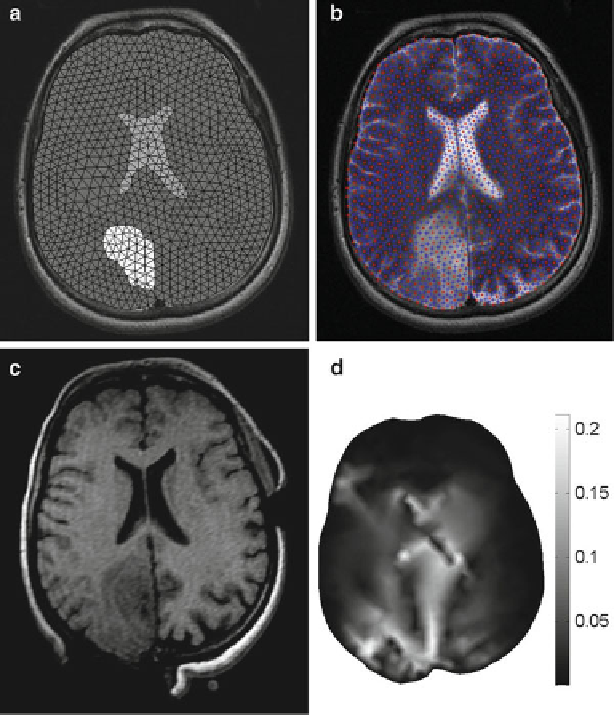
Fig. 3 Comparison of simulation results obtained from FE model and statistical meshless model
(a) Statistic meshless model with 755 nodes, 1,417 integration points, intracranial area is modeled
as a whole, without explicitly separating the tumor and ventricles (b) Finite Element model with
817 nodes, 1,541 elements, three parts are modeled separately: parenchyma, tumor and ventricle.
(c) Intraoperative MRI (with craniotomy location shown). (d) Difference in computed deformation
field from the two models (unit: mm)
constitutes a significant problembecause of complexity of the brain-skull interface. As
the skull is orders of magnitude stiffer than the brain tissue, we assumed it to be rigid. A
very efficient and realistic contact algorithmdeveloped in [
24
] was employed tomodel
this interaction as a finite sliding, frictionless contact. To reduce the computation time
required for stable solution with the prescribed accuracy, dynamic relaxation [
25
]was
used for estimating the parameters as well as the termination criteria.
The difference in computed deformation fields from the two models is shown in
Fig.
3
. For each node in the finite element model, the displacement was compared to