Biomedical Engineering Reference
In-Depth Information
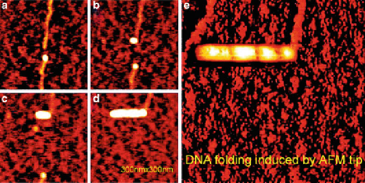
Fig. 5.2
A series of AFM images indicating that DNA strands were fold up by an AFM tip. (
a
-
d
)
AFM images indicating a cutting and folding process of a DNA strand. (
e
) A DNA strand was
folded up to form a thick rod. Image size: 300 nm
300 nm (Reprinted with the permission from
Ref. [
13
]. Copyright 2002 American Chemical Society)
dsDNA [
15
]. This is supported by the fact that the time requested to break ssDNA
is shorter than that of dsDNA in the same dissection procedure under the same load.
This result implies that dsDNA and ssDNA have different mechanical properties.
5.3
Isolating and Analyzing Single DNA Fragments
by AFM Nanomanipulation
The idea to isolate a particular biomolecule from a surface with an AFM tip can be
dated back to more than a decade ago. Heckl et al. have demonstrated dissection
and subsequent extraction of genetic materials from chromosomes [
16
]. Xu and
Ikai have found that AFM tips could occasionally pick up plasmid DNA from a
solid surface [
17
]. Guthold et al. have tried to use AFM to isolate single aptamer
molecules [
18
]. However, the approaches mentioned above are not adequate to
realize the positional isolation which is required for practical applications such as
direct molecular haplotyping of a genomic DNA.
Recently, we have developed a general protocol for isolation of individual DNA
fragments by using a special AFM nanomanipulation technique [
19
]. The isolation
procedure was carried out by real-time switching of the AFM operation from
tapping mode (for imaging and positioning) to contact mode (for isolation). The
isolation result could be revealed by scanning the same area again with tapping
mode AFM. A typical example for isolating single DNA fragment after AFM
cutting is shown in Fig.
5.3
. After the confirmation of the pickup of the target
Search WWH ::

Custom Search