Biomedical Engineering Reference
In-Depth Information
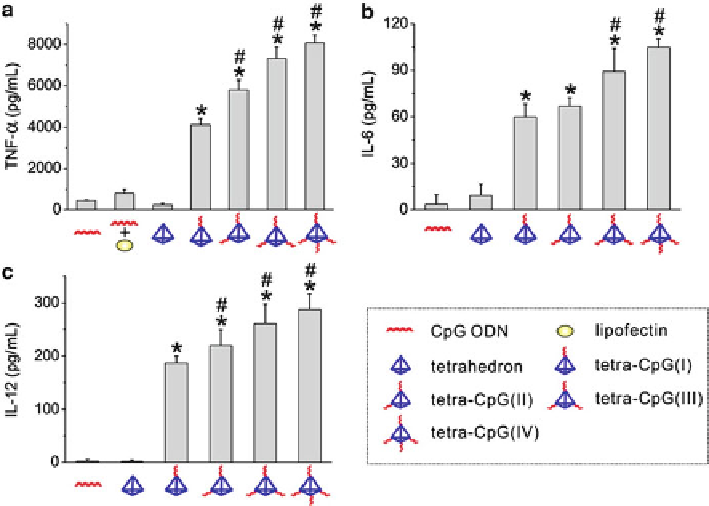
Fig. 15.5
Cytokine release from RAW264.7 cells stimulated by DNA nanostructures. Comparison
of (
a
)TNF-R,(
b
)IL-6,and(
c
) IL-12 release stimulated by CpG ODN (80 nM) and DNA
nanostructures of 20 nM. Error bars represent standard deviation (
SD
) of at least three independent
measurements. *
P
0.05
significantly different from tetra-CpG(I) (Reprinted with permission from Ref. [
11
]. Copyright
2011 American Chemical Society)
<
0.001 significantly different from CpG ODN and tetrahedron. #
P
<
aid of transfection agents, and once inside the cell, the functional DNA tetrahedrons
were most located at the cytoplasm. And the DNA tetrahedron structures could
remain intact for at least several hours both in 50% non-inactivated fetal bovine
serum (FBS) and in cells.
By studying the immunostimulatory effects of the functional CpG-DNA tetra-
hedron nanostructures using ELISA assays, they found that by activating Toll-like
receptor 9 (TLR9) and then the downstream pathway, all functional CpG-DNA
tetrahedron nanostructures dramatically induced the production of various proin-
flammatory cytokines including tumor necrosis factor (TNF)-R, interleukin (IL)-6,
and IL-12. Due to the greatly enhanced cellular uptake efficiency of the functional
CpG-DNA tetrahedron nanostructures as compared to single-stranded DNA, the
level of immunostimulatory effects were increased by 9-18 times comparing to free
CpG oligodeoxynucleotides (Fig.
15.5
,Ref.[
11
]).
The evaluation of the cytotoxicity of the functional CpG-DNA tetrahedron
nanostructures by standard colorimetric MTT (3-(4,5-dimethylthiazol- 2-yl)-2,5-
diphenyl tetrazolium bromide) assay revealed that the functional CpG-DNA tetra-
hedron did not induce measurable loss in the viability of cells even at a concentration
Search WWH ::

Custom Search