Biomedical Engineering Reference
In-Depth Information
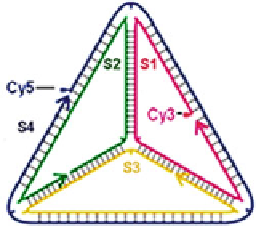
Fig. 15.2
Schematic
showing the design of the
DNA tetrahedrons (Reprinted
with permission from Ref.
[
10
]. Copyright 2011
American Chemical Society)
tetrahedron structure they used has edges of 7 nm each, which was originally
designed by Goodman et al. [
8
,
9
], after tests with specific and nonspecific DNA
nucleases, and comparing with the results of DNA single strands, they found that
the DNA tetrahedron structure has superior stability over DNA single strands against
enzymatic digestions. More surprisingly, they found that in 10% fetal bovine serum
(consisting complex mixture of nucleases and other proteins), the DNA tetrahedron
structure can be stable for 42 h, comparing to only 0.8 h for DNA single strands. This
experiment brings the hope that DNA tetrahedron nanostructures might be stable in
cells and might be used for drug delivery applications.
Turberfield's group [
10
] then investigated the ability of this enzyme-tested DNA
tetrahedron nanostructure to enter live cultured mammalian cells and its potential as
a drug delivery nanocarrier.
They covalently attached organic fluorescence dye Cy5 to one of the four 63-
base DNA strands of the DNA tetrahedron (Fig.
15.2
,Ref.[
10
]), Cy3 was later to
the designed position of another DNA strands to test structure integrity of this DNA
tetrahedron.
By comparing transfection levels of Cy5-labeled DNA tetrahedron inside plated
human embryonic kidney cells with controlled single-stranded DNA, also by
comparing transfection levels of Cy5-labeled DNA tetrahedron with and without the
addition of the cationic lipid transfection reagent Lipofectin (Fig.
15.3
,Ref.[
10
]),
their confocal microscopy and flow cytometry results showed that DNA tetrahedron
structure could easily enter cells alone, probably because of its size and compact
structure. Organelle-specific dye (Hoechst 34580 and LysoSensor Green) stain
experiment showed that transfected tetrahedra are clearly locating at the cytoplasm.
And fluorescence resonance energy transfer (FRET) experiment was performed
to test the structure stability of this DNA tetrahedron structure after transfection,
using a modified design with two fluorescence dyes, Cy5 and Cy3, in close vicinity
(Fig.
15.2
); FRET results showed that the DNA tetrahedron structure remains intact
in cells even 48 h after transfection.
The high transfection levels of the DNA tetrahedron nanostructure, and its good
structure stability inside the cells, indicated that the DNA tetrahedron structure
could be a promising drug delivery nanocarrier.
Search WWH ::

Custom Search