Biomedical Engineering Reference
In-Depth Information
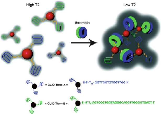
Fig. 13.10
MRI-based protein sensing based on aptamer-functionalized CLIO nanoparticles. The
CLIO nanoparticles modified with two different thrombin aptamers (
green
and
blue
) assemble in
the presence of thrombin, resulting in decreased T2 value and darker MR images (Reprinted with
the permission from Ref. [
145
]. Copyright 2008 American Chemical Society) (Color figure online)
Recently the Lu group developed a general strategy to construct smart MRI
agents for detecting chemical and biological molecules based on aptamer-
functionalized CLIO nanoparticles. Adenosine aptamer-linked CLIO nanoparticle
aggregates could be disassembled in the presence of adenosine, which was observed
by MRI with a dramatic enhancement in the brightness of T2-weighted MR
images [
144
]. This MRI enhancement was highly selective for adenosine, as other
nucleobases did not result in any change in MR images. In order to demonstrate the
design of smart MRI contrast agents that respond to proteins instead of small
molecules, the Lu group has developed the MRI system for thrombin [
145
].
Due to multiple aptamer-binding sites in each thrombin molecule, the addition of
thrombin could trigger the assembly of the thrombin aptamer-functionalized CLIO
nanoparticles into an aggregated structure, which could be observed by the decrease
of brightness in T2-weighted MR images (Fig.
13.10
). The system demonstrated
is specific to thrombin and shows a detectable change in MRI signal with 25 nM
thrombin in human serum. Based on a similar design, an MRI-based sensor for
lysozyme protein detection was achieved using CLIO nanoparticles conjugated
with lysozyme aptamer [
146
]. Since such an MRI-based detection method is light-
independent, without background interference by scattering or autofluorescence, it
may be suitable for noninvasive in vivo detection of small molecular markers.
Search WWH ::

Custom Search