Biomedical Engineering Reference
In-Depth Information
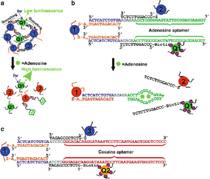
Fig. 13.7
Multiplexed sensor based on aptamer-linked QDs and AuNPs. (
a
)AuNP1,2,and
quantum dot Q1 were assembled by an adenosine aptamer with quenched QD emission. Addition
of adenosine disassembled the aggregates and recovered the QD emission. DNA sequences and
NPs linkages for the adenosine sensor (
b
) and for the cocaine sensor (
c
) (Reprinted with the
permission from Ref. [
112
]. Copyright 2007 American Chemical Society)
quadruplex conformation of the aptamer was stabilized due to the binding with
thrombin, resulting in the displacement of the quencher strand and a concomitant
enhancement in fluorescence. Strano and coworkers developed a different type of
QD-aptamer sensor based on the charge transfer [
111
]. In this work, PbS QDs were
functionalized with thrombin aptamers. They found that the interaction of thrombin
with aptamers brought them into close proximity with the QDs and thus could
quench the fluorescence of QDs due to the charge transfer from thrombin to the
QDs. The sensor showed a detection limit of 1 nM and high selectivity.
Because of the highly tunable emission properties of QDs, the Lu group reported
a multiplex sensor for detection of adenosine and cocaine using aptamers conjugated
with QDs and AuNPs (Fig.
13.7
)[
112
]. The aggregates of QDs Q1 (emission at
525 nm) and AuNPs were formed by the adenosine aptamer linker, resulting in
the quenched QD emission because of energy transfer to nearby AuNPs. Addition
of adenosine disassembled the aggregates and thus increased emission intensity
Search WWH ::

Custom Search