Biomedical Engineering Reference
In-Depth Information
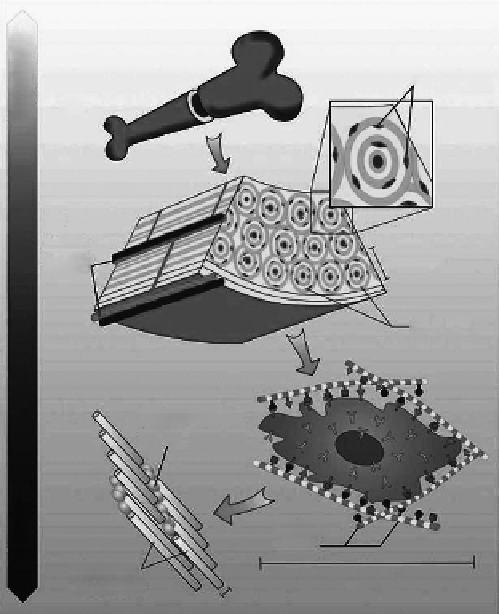
Osteocytes
(a)
(b)
Blood
vessels
100-500 μm
Osteons
(c)
(d)
Hydroxyapatite
crystals
Collagen
fibers
Collagen
molecules
10-50 μm
1nm
Figure 9.30
Hierarchical organization of bone over different length scales. Bone has a strong calcified outer compact layer (a),
which comprises many cylindrical Haversian systems, or osteons (b). The resident cells are coated in a forest of
cell membrane receptors that respond to specific binding sites (c) and the well-defined nanoarchitecture of the
surrounding ECM (d). (From Stevens, M. M. and George, J. H. 2005.
Science
310: 1135-1138. With permission.)
osteoblasts (improve bone forming) and osteoclasts (improve bone resorbing) [158,159].
Most research pay attention to the osteoblast response to chitosan films or scaffolds.
Chitosan was shown to be an effective substrate that supports the initial attachment and
the spreading of osteoblasts. The viability of the attached osteoblasts on chitosan sub-
strates generally increased significantly with an increase in the cell seeding time. Osteoblast
cells are not fully attached on the surfaces of the chitosan substrates at 2 h after cell seed-
ing and they are round. At 8 h after cell seeding, osteoblasts on the chitosan film counter-
parts assumed a spindle shape [160]. Osteoblasts displayed phenotypes that were well
spread with a developed cytoskeleton [161]. Moreover, chitosan supports osteoblasts to
produce mineralized tissues [162]. Takakuda and coworkers [163] found that chitin-chito-
san can be used as a bone formation accelerator. After the 2-week implantation of chitin-
chitosan coating on PLA fiber into holes drilled into the distal metaphyses of rat femora,
the mean area of bone tissue in the experimental group was 0.532 ± 0.145 mm
2
, while that
in the control group was 0.113 ± 0.049 mm
2
. The success of an implant not only depends on
the interaction with osteoblasts. Osteoclasts also play a very important role in the forma-
tion process of new bone. Cartmell SH and coworkers [164] found that chitosan supported
osteoclast culture, but the proliferation capacity of osteoclasts is lower than that of osteo-
blasts on chitosan film. Chitosan recorded significantly higher amounts of DNA in the
Search WWH ::

Custom Search