Biomedical Engineering Reference
In-Depth Information
TAble 9.3
The Cell Cycle of Fibroblasts on the Scaffold
Cell Cycle (%)
Time (Days)
G
0
/G
1
S
G
2
/M
Cell Death (%)
3
55.4
6.96
36.2
1.61
7
51.5
7.62
39.1
2.01
14
50.4
5.86
40.8
3.14
Source:
From Sun, L. P. et al. 2009.
Biomed Mater
4: 055008. With permission.
scaffolds. Moreover, the chitosan-collagen scaffolds with 30 wt% chitosan showed
the most significant increase in relative cell viability [108]. Most fibroblasts seeded on the
scaffolds come in the normal cell cycle. The cell death rate is very low (
cf.
Table 9.3). The
percentages of fibroblasts cultured in the dish for 3 days in the G
0
/G
1
, S, and G
2
/M phase
cells and apoptotic cells are 57.8%, 37.8%, 4.4%, and 0.5%, respectively. The S-phase cells
cultured in the chitosan-collagen scaffold decrease apparently and the G
2
/M phase cells
increase remarkably, which demonstrates that the collagen-chitosan scaffolds promote
the S-G
2
/M phase transition [109]. However, fibroblasts may locate at the peripheral area
of the chitosan-based scaffold and cover some pores of the scaffold surface, which may
prevent nutrition supply, limit cell growth, and result in more inhomogeneous tissues. In
order to decrease these phenomena, Tan and coworkers [110] seeded human dermal fibro-
blasts into the chitosan-collagen scaffold using a flow perfusion system. It was found that
seeding at the perfusion rate of 0.125 mL/min and 0.25 mL/min could achieve signifi-
cantly higher seeding efficiencies than that at 0.5 mL/min. In comparison with the static
seeding method, the perfusion seeding at 0.25 mL/min promoted a more efficient cell uti-
lization and achieved higher initial cell densities without compromising the uniformity of
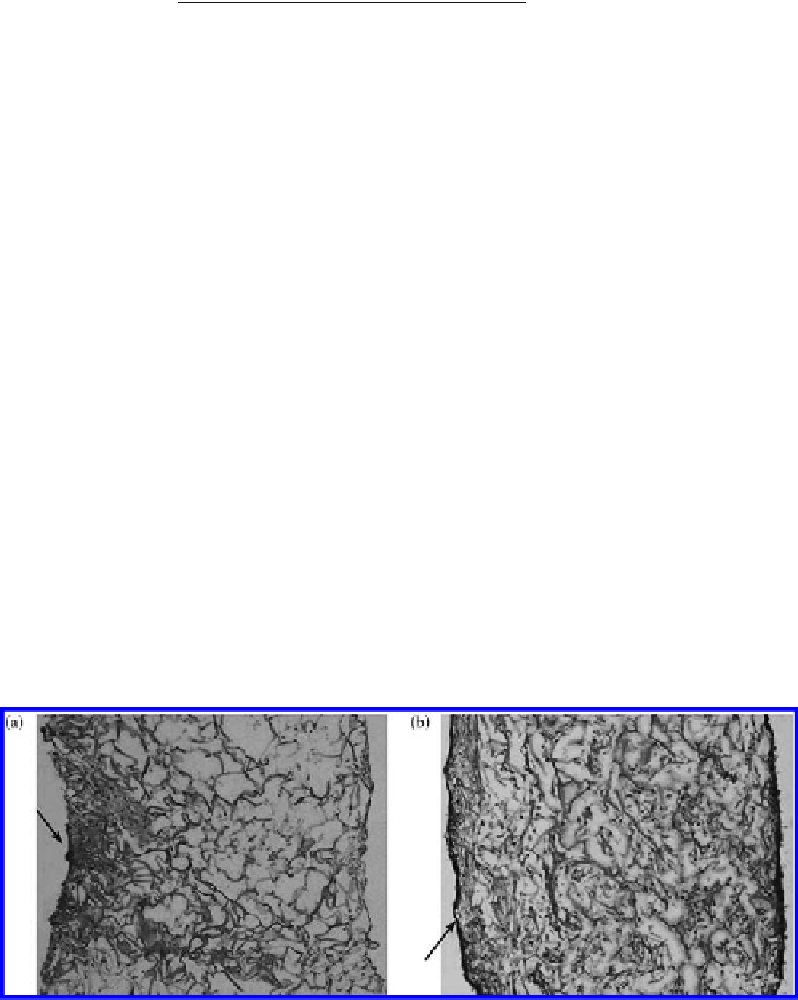
initial cell distribution. The uniform distribution of cells achieved using perfusion seeding
was found to be favorable to prolong cell proliferation period, increase the number of cells,
and form more homogeneous morphology of constructed tissues, as shown in Figure 9.20.
In addition, fibroblasts cultured under mechanical stimulation (subjected to the cyclic
Figure 9.20
Cell distribution on the cross section of the 4-week chitosan-collagen constructs. Hematoxylin and eosin
(H&E)-stained cross section of static cultured 4-week constructs seeded by the static (a) and perfusion (b) meth-
ods (the arrow denotes the top surface of constructs). (From Ding, C. M. et al. 2008.
Proc Biochem
43: 287-296.
With permission.)


Search WWH ::

Custom Search